. 2023 Jun 14:2023:8818561.
doi: 10.1155/2023/8818561. eCollection 2023.

The Effect of Distraction Osteogenesis on Peripheral Nerve Regeneration in Rats: A Preliminary Study In Vivo

Affiliations

The Effect of Distraction Osteogenesis on Peripheral Nerve Regeneration in Rats: A Preliminary Study In Vivo

Kai Liu et al. J Tissue Eng Regen Med. .

Abstract

Distraction osteogenesis (DO) is a widely employed method for the treatment of limb discrepancies and deformity correction. This study aimed at observing the histomorphological and ultrastructural changes of peripheral nerves around the distraction area during DO and investigating the self-repair mechanism of peripheral nerves in a rat DO model. Sixty rats underwent right femoral DO surgery and were randomly separated into six groups: Control (latency, no distraction, n = 10), Group 0-week (after distraction, n = 10), Group 2-week (n = 10), Group 4-week (n = 10), Group 6-week (n = 10), and Group 8-week (n = 10) at consolidation phase. The right femur of rats in Group 0-week, Group 2-week, Group 4-week, Group 6-week, and Group 8-week was subjected to continuous osteogenesis distraction at a rate of 0.5 mm/day for 10 days. Motor nerve conduction velocity (MNCV) of the sciatic nerve, sciatic function index (SFI), histological analyses, and transmission electron microscopy were conducted to evaluate nerve function. The MNCV and SFI of Group 0-week, Group 2-week, Group 4-week, and Group 6-week were significantly lower than the Control (P < 0.05). No statistical differences were found between the Control and Group 8-week in terms of MNCV and SFI (P > 0.05). Injuries to nerve fibres and nodes of Ranvier were observed in the Group 0-week, whereas the nerve fibres returned to the normal arrangement in the Group 8-week and oedema of myelin disappeared, with the continuity of axons and lamellar structure of myelin being restored. Femoral DO in rats with a rate of 0.5 mm/day may cause sciatic neurapraxia, which can be self-repaired after 8 weeks of consolidation. The paraneurium around the sciatic nerve enables it to glide during the distraction phase to reduce the occurrence of injurious changes.


Conflict of interest statement

The authors declare that they have no conflicts of interest.

Figures
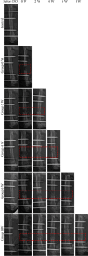
Figure 1
Figure 1
Radiographs of rat femur after DO surgery using external fixation (n = 10 per group).
Figure 2
Figure 2
Results of electrophysiology and SFI in different groups (n = 10 per group). (a) The motor nerve conduction velocity (MNCV) of the Group 0-week was significantly lower than the other groups (P < 0.05), and MNCV increased with increasing duration of consolidation. (b) The sciatic function index (SFI) of the Group 0-week was significantly lower than the other groups (P < 0.05), and the absolute value of SFI decreased with the increasing duration of consolidation.
Figure 3
Figure 3
Histological results of the sciatic nerves in different groups (HE staining, n = 3 per group). (a) Wavy running nerve fibres were observed in the control, as characterized by the obvious nodes of Ranvier (red arrow) and scattered distribution in Schwann's cell nuclei (yellow arrow). (b) Disorganized nerve fibres were observed in Group 0-week with the widened distance between some nerve bundles (red arrows) and medullary spheroid changes appearing to be a lipid droplet-like structure (yellow arrows). (c) Mild reparative changes were seen in Group 2-week with still-persisting lipid droplet-like structures (yellow arrows). (d) Self-repaired nerve fibres were observed in Group 4-week with myeloid-like changes still present (yellow arrows). (e, f) At Group -week and Group 8-week, the nerve fibres had recovered to the normal arrangement, with the clear observation of the nerve axon, the node of Ranvier, and internode structures.
Figure 4
Figure 4
Transmission electron microscopy results of the sciatic nerves in different groups (n = 2 per group). In the Control, closely arranged lamellar myelin structures (red arrows) were observed with the axonal membrane being smooth and intact containing microtubule microfilaments. At Group 0-week, the myelin lamellae of myelinated nerve fibres were loosely sheeted apart (yellow arrows), with oedema of Schwann cells, vacuolar myelin degeneration, and even compression of axons also visible. Mild reparative changes and new myelination occurred in Group 2-week (yellow arrows). Active proliferation of Schwann cells was identified in Group 4-week, although degenerated nerve fibres were still present (yellow arrows). At Group 6-week and Group 8-week, the arrangement of nerve fibres gradually returned to normal (yellow arrow), with a large number of newly formed myelin sheaths observed and occasionally with myelosphere changes in myelinated nerve fibres (red arrows).
Figure 5
Figure 5
Immunohistochemistry images of p38MAPK in different groups (n = 5 per group). (a, b) The semiquantitative analysis indicated that p38MAPK was significantly expressed in Group 2-week, Group 4-week, and Group 6-week compared to the Control (black arrows, P < 0.05). However, the expression of p38MAPK was downregulated in the Group 8-week, and there was no statistically significant difference compared to the control group.

References

    1. Balci H. O., Bayram S., Pehlivanoglu T., Anarat F. B., Eralp L., Şen C. Effect of lengthening speed on the quality of callus and complications in patients with congenital pseudarthrosis of tibia. International Orthopaedics . 2021;45:1517–1522. doi: 10.1007/s00264-021-05011-7. - DOI - PubMed
    1. Hosny G. A. Limb lengthening history, evolution, complications and current concepts. Journal of Orthopaedics and Traumatology . 2020;21 doi: 10.1186/s10195-019-0541-3. - DOI - PMC - PubMed
    1. Liu Y., Cai F., Liu K., Liu J., Zhang X., Yusufu A. Cyclic distraction–compression dynamization technique enhances the bone formation during distraction osteogenesis. Frontiers in Bioengineering and Biotechnology . 2022a;9 doi: 10.3389/fbioe.2021.810723. - DOI - PMC - PubMed
    1. Xu J., Sun Y., Wu T., Liu Y., Shi L., Zhang J. Enhancement of bone regeneration with the accordion technique via HIF‐1α/VEGF activation in a rat distraction osteogenesis model. J Tissue Eng Regen M . 2018;12 doi: 10.1002/term.2534. - DOI - PubMed
    1. Liu Z., Liu Y., Yushan M., Yusufu A. Enhanced nerve regeneration by bionic conductive nerve scaffold under electrical stimulation. Frontiers in Neuroscience . 2022b;16 doi: 10.3389/fnins.2022.810676.810676 - DOI - PMC - PubMed

Publication types

LinkOut - more resources