Review
. 2024 Oct 11:49:135-155.
doi: 10.1016/j.jot.2024.08.003. eCollection 2024 Nov.

Porous metal materials for applications in orthopedic field: A review on mechanisms in bone healing

Affiliations
Review

Porous metal materials for applications in orthopedic field: A review on mechanisms in bone healing

Yutong Ma et al. J Orthop Translat. .

Abstract

Background: Porous metal materials have been widely studied for applications in orthopedic field, owing to their excellent features and properties in bone healing. Porous metal materials with different compositions, manufacturing methods, and porosities have been developed. Whereas, the systematic mechanisms on how porous metal materials promote bone healing still remain unclear.

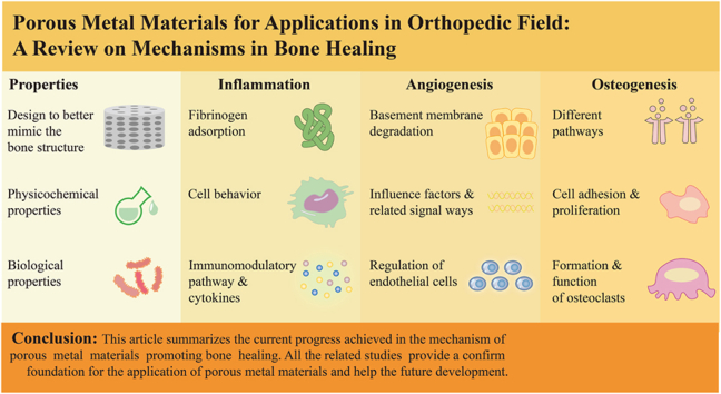
Methods: This review is concerned on the porous metal materials from three aspects with accounts of specific mechanisms, inflammatory regulation, angiogenesis and osteogenesis. We place great emphasis on different cells regulated by porous metal materials, including mesenchymal stem cells (MSCs), macrophages, endothelial cells (ECs), etc.

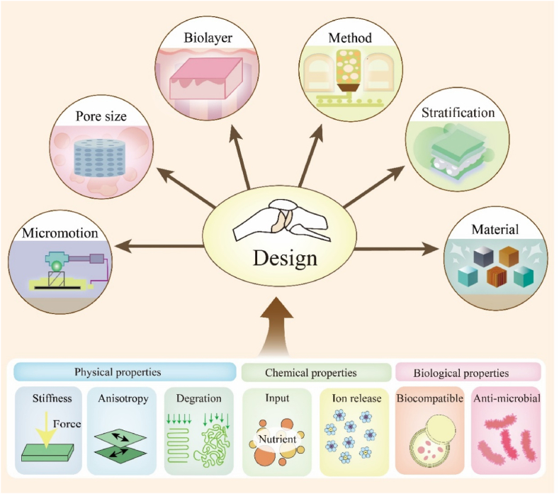
Result: The design of porous metal materials is diversified, with its varying pore sizes, porosity material types, modification methods and coatings help researchers create the most experimentally suitable and clinically effective scaffolds. Related signal pathways presented from different functions showed that porous metal materials could change the behavior of cells and the amount of cytokines, achieving good influence on osteogenesis.

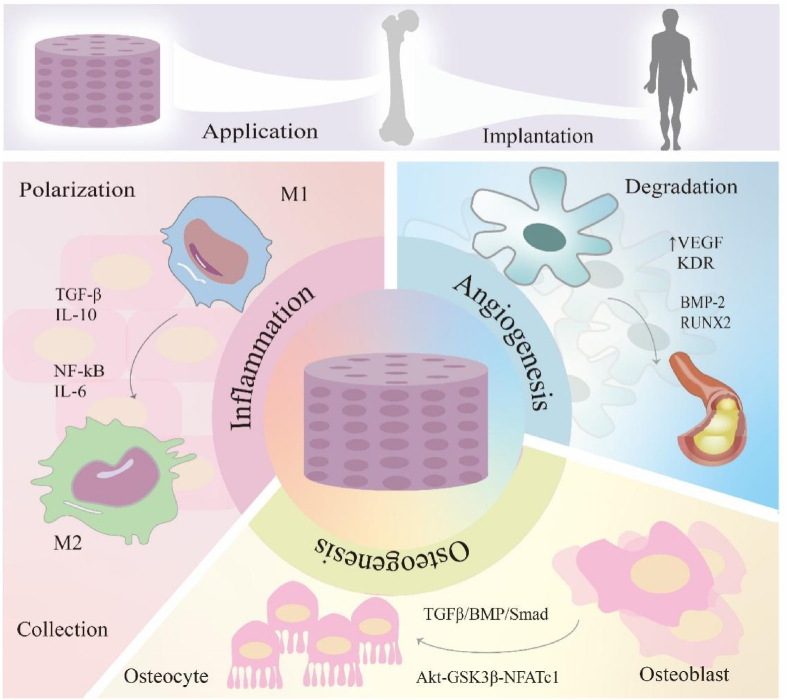
Conclusion: This article summarizes the current progress achieved in the mechanism of porous metal materials promoting bone healing. By modulating the cellular behavior and physiological status of a spectrum of cellular constituents, such as macrophages, osteoblasts, and osteoclasts, porous metal materials are capable of activating different pathways and releasing regulatory factors, thus exerting pivotal influence on improving the bone healing effect.

The translational potential of this article: Porous metal materials play a vital role in the treatment of bone defects. Unfortunately, although an increasing number of studies have been concentrated on the effect of porous metal materials on osteogenesis-related cells, the comprehensive regulation of porous metal materials on the host cell functions during bone regeneration and the related intrinsic mechanisms remain unclear. This review summarizes different design methods for porous metal materials to fabricate the most suitable scaffolds for bone remodeling, and systematically reviews the corresponding mechanisms on inflammation, angiogenesis and osteogenesis of porous metal materials. This review can provide more theoretical framework and innovative optimization for the application of porous metal materials in orthopedics, dentistry, and other areas, thereby advancing their clinical utility and efficacy.

Keywords: Angiogenesis; Bone regeneration; Immunomodulation; Osteogenesis; Porous metal materials.


Conflict of interest statement

All authors declare that there are no competing interests.

Figures

Image 1
Graphical abstract
Figure 1
Figure 1
Applications of porous metal material in bone cause different kinds of reactions. Inflammation: the usage of porous metal material activates the macrophage from M1 to M2, through NF-kB and IL-6 factors. It also promotes the collection of mesenchymal stem cells. Angiogenesis: through the process of angiogenesis, porous metal material improves the basement membrane degradation by activating the amount of VEGF, KDR, BMP-2 and RUNX2. Osteogenesis: the osteoblasts are activated into osteocyte through binding of signal ways.
Figure 2
Figure 2
The design of porous metal materials. The design of porous metal materials is apparently important, including the pore size, biolayers, material category, manufacturing methods, stratification interface micromotion, topographical design, and so on. The aim of proper design is to make porous materials with excellent physical, chemical and biological properties, including stiffness, anisotropy, degradation, nutrition input, ion release, biocompatibility and anti-bacteria.
Figure 3
Figure 3
Different features of porous metal materials. A Digital photographs of metal scaffolds of low porosity (LP), high porosity (HP) and zero porosity (ZP) and their respective SEM images. B Antibacterial properties of scaffolds of ZP, LP, and HP cultured with S. aureus for 24 h. C Mechanical performance of scaffolds, including stress–strain curves at 2 % strain rate, compressive strength, rule-of-Mixtures model and the fit data, and the porosity of the respective scaffolds. D XRD of corroded samples. E MTT assay of pre-osteoblast cell viability cultured in 10 % dilution extracts prepared by incubation of samples with cell media for up to 5 days. F Corrosion rate taken from mass loss after immersion in Hank's solution for 1 month. G EDS of respective images. Reprinted with permission from Ref. [25]. © 2020 Elsevier B.V.
Figure 4
Figure 4
Porous metal materials regulate the inflammatory phase. A: The SEM image of fibrinogen and magnesium combination biomaterials modulating macrophage phenotype. Magnification:250×. Scale:400 μm. B: Effect of macrophage secretome on MSC osteogenic differentiation. MSC were incubated for 14 days with biomaterial extracts (extract CTRL=Fg, Mg and FgMg biomaterials extracts) or the secretome of macrophages unstimulated (M0), or LPS-IFNγ stimulated (M1), in presence/absence of biomaterial extracts (CTRL = conditioned media from macrophages not exposed to extracts; Fg, Mg and FgMg extracts = conditioned media from macrophages preconditioned with Fg, Mg and FgMg extracts alone), as indicated. Basal and osteogenic (osteo) media were used as controls. ALP and Alizarin Red staining were performed. C: Schematic representation of the cross-talks among MΦs to OBs and ECs. Signaling pathways of MΦs to OBs involved in the present study. Signaling pathways of MΦs to ECs involved in the present study. Cross-talks among MΦs, OBs, and ECs. D: Immune response of the modified surface and the interaction of bone immunoregulation, osteogenesis, and angiogenesis. FE-SEM images of MΦs morphology on samples at different magnifications. E: The effect of surface nano-microscopic characteristics on cytokines and interaction of bone immunoregulation, osteogenesis and angiogenesis, including the expression of OBs (BMP2, COL1, OCN, OPG, TGF-β1, VEGFA, ALP, OSX, Runx2 and SMAD1/4/5/8) osteogenic genes and genes related to angiogenesis in ECs (eNOS, VEGF and BMP2). Reprinted with permission from Refs. [85,115]. © 2020 Acta Materialia Inc. Published by Elsevier Ltd. All rights reserved. © 2018 Acta Materialia Inc. Published by Elsevier Ltd. All rights reserved.
Figure 5
Figure 5
Implantation of porous metal materials activates a variety of signal pathways and cytokines related to angiogenesis. A Hypoxia occurs under the use of porous metal materials usually causing upregulation of HIF-a, thereby enhancing the expression of related genes. B Porous metal materials induce the angiogenesis of HUVECs via FGF signaling pathway. It acts on FGFR by promoting the expression of FGF to promote the activation of intracellular related cellular pathways, including RAS-MAPK and PI3K-AKT pathways [143]. C The first step of angiogenesis is the degradation of basement, while the release of related cytokines contributes to the proliferation of endothelial. Finally, these factors contribute to the angiogenesis.
Figure 6
Figure 6
Influence of porous metal materials on osteogenesis. The degradation of porous metal materials causes the dispersed distribution of metal ions, which causes the converge of osteoblast. BMP enters the osteoblast and makes the phosphorylation of Smad, which inhibits the expression of RUNX2 and up-regulates ALPL. The phosphorylation of GSK3β triggers β-linked catenin, then promotes the expression of Wnt, which finally promote the expression of RUNX2. It also improves the AMPK signal pathway, which improves OGN, OGP, ALP, OSX, OSN and OCN, linking to osteogenesis. The activation of AMPK also inhibits mevalonate, promoting the differentiation of osteoblast.
Figure 7
Figure 7
Porous metal materials act important part in osteogenesis. A Scanning electron microscopy was used to observe the pore size of the two materials, and the energy spectrum was used to analyze the surface composition of the material. B Adhesion of BMSCs. Light microscopy image of BMSCs and fluorescence microscopy image of live/dead cell staining of BMSCs cocultured with the porous tantalum and pTi6Al4V for 1 day. C Q-PCR showed p-ERK and ERK protein expression of BMSCs cocultured with pTa and pTi6Al4V for 7, 14 and 21 days, GAPDH as an internal reference. D SEM images and EDX elemental maps of the bone sections with 20Fe@40Zn scaffolds after 3 months of implantation. They showed new bone and connective tissue layer surrounding the skeletons of the scaffold. E Histological analysis of the bone tissue around the porous 20Fe@40Zn scaffolds. F Quantified BV/TV and BIC from the micro-CT reconstructions. Reprinted with permission from Refs. [183,187] © 2019 The Authors. Published by Elsevier (Singapore) Pte Ltd on behalf of Chinese Speaking Orthopedic Society. © 2020 Acta Materialia Inc. Published by Elsevier Ltd. All rights reserved.

Similar articles

Cited by

References

    1. Hamiti Y., Yushan M., Lu C., Yusufu A. Reconstruction of massive tibial defect caused by osteomyelitis using induced membrane followed by trifocal bone transport technique: a retrospective study and our experience. BMC Surg. 2021;21(1):419. - PMC - PubMed
    1. Chen W., Fang R. [Internal fixation of mandibular comminuted fracture with mini-titanium plate: a retrospective study of 21 cases] Shang Hai Kou Qiang Yi Xue. 2020;29(3):333–336. - PubMed
    1. Won A.M., Montgomery P., Aponte-Wesson R., Chambers M. Implant-supported and magnet-retained oral-nasal combination prosthesis in a patient with a total rhinectomy and partial maxillectomy due to cancer: a clinical report. J Prosthet Dent. 2017;117(2):315–320. - PubMed
    1. Li D., Li Y., Shrestha A., Wang S., Wu Q., Li L., et al. Effects of programmed local delivery from a micro/nano-hierarchical surface on titanium implant on infection clearance and osteogenic induction in an infected bone defect. Adv Healthcare Mater. 2019;8(11) - PubMed
    1. Chang C., Greenspan A., Gershwin M.E. The pathogenesis, diagnosis and clinical manifestations of steroid-induced osteonecrosis. J Autoimmun. 2020;110 - PubMed

LinkOut - more resources