. 2025 Aug;48(4):961-978.
doi: 10.1007/s13402-025-01057-6. Epub 2025 Apr 14.

THEMIS2 contributes to ovarian cancer metastasis via DOCK4-mediated activation of Rap1 signaling

Affiliations

THEMIS2 contributes to ovarian cancer metastasis via DOCK4-mediated activation of Rap1 signaling

Kaixia Zhou et al. Cell Oncol (Dordr). 2025 Aug.

Abstract

Purpose: Ovarian cancer (OC) is the most lethal gynecological malignancy, with widespread metastasis and ascites being the leading causes of patient mortality. However, the mechanisms driving OC metastasis have not been sufficiently studied. This study aimed to investigate the mechanisms and key molecules promoting OC metastasis.

Methods: Public databases (StemChecker, GeneCards, GEO, and TCGA) were screened to identify metastasis-associated genes. Immunohistochemical staining and western blotting were employed to evaluate THEMIS2 expression and epithelial-mesenchymal transition (EMT) marker profiles across experimental groups. RNA sequencing coupled with pathway enrichment analysis revealed THEMIS2-regulated signaling pathways, while immunoprecipitation-mass spectrometry was utilized to identify THEMIS2 interaction partners. GST pull-down assays for active Rap1 quantified Rap1-GTP levels under varying THEMIS2 expression conditions. Wound healing and transwell invasion assays respectively assessed migratory and invasive capacities of OC cells following THEMIS2 expression perturbations in vitro. Abdominal cavity implantation metastasis model was established to evaluate OC cell colonization and invasive potential in vivo.

Results: THEMIS2 expression is significantly elevated in OC tissues compared to normal ovarian tissues, and its high expression correlates with poor prognosis and malignant features. Experimental manipulation of THEMIS2 levels revealed that knockdown impended the migratory and invasive capacities of OC cells both in vitro and in vivo, while its overexpression exacerbated metastasis. THEMIS2 is involved in EMT and cytoskeleton rearrangement. RNA-seq analysis revealed that THEMIS2 positively correlates with Rap1 signaling pathway. Inhibition of Rap1 activity reversed the metastasis-promoting effects induced by THEMIS2 overexpression both in vitro and in vivo. Mechanistically, we uncovered that THEMIS2 functions as a molecular scaffold that recruits TBK1 (TANK Binding Kinase 1) to DOCK4 (Dedicator of Cytokinesis 4), facilitating site-specific phosphorylation at serine 1787 (S1787). This post-translational modification enables DOCK4 to engage with CRKII, subsequently triggering Rap1 signaling activation. These findings suggest that THEMIS2 promotes the metastatic potential of OC cells via DOCK4-mediated activation of Rap1 signaling.

Conclusion: THEMIS2 may serve as a predictive biomarker for OC prognosis, and targeting the Rap1 signaling pathway with specific inhibitors represents a promising therapeutic strategy for OC treatment.

Keywords: DOCK4; Ovarian cancer; Rap1 signaling; THEMIS2.


Conflict of interest statement

Declarations. Ethics approval and consent to participate: This study was conducted in compliance with the Declaration of Helsinki principles. Informed consents were obtained from all the subjects. Ethics approval for human subjects was provided by the Ethics Committee of Fudan University Shanghai Cancer Center. Ethics approval for animal work was provided by the Institutional Animal Care and Use Committee of Shanghai Cancer Center. Consent for publication: Not applicable. Competing interests: The authors declare no competing interests.

Figures
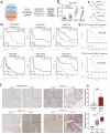
Fig. 1
Fig. 1
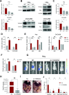
The clinical relevance of THEMIS2 in ovarian cancer. (A) Work flow of THEMIS2 selection. SRGs: Stemness-related genes; IRGs: Invasion-related genes. (B) NFKBIB, ZNF70 EPB41 and THEMIS2 expression analysis in primary and metastatic OC tissue using GSE222982 cohort. (C) Survival curves for patients with OC based on the expression of EPB41 or THEMIS2 from TCGA data. (D) Kaplan-Meier analysis of PFS of OC patients according to THEMIS2 mRNA expression, generated based on GEO cohorts (GSE19829, GSE30161, GSE3149, GSE9891, GSE15622 and GSE27651 datasets). (E) Kaplan-Meier plot of the correlation between THEMIS2 expression and patient survival using TCGA Ovarian Proteome datasets. (F) Representative immunohistochemical images of THEMIS2 expression in OC and normal tissues. Tumor -/-: Patients suffered neither lymphatic metastasis nor ascites; Tumor -/+: Patients suffered either lymphatic metastasis or ascites; Tumor +/+: Patients suffered lymphatic metastasis and ascites. (G) Top: The relative positive signal of THEMIS2 in OC and normal ovarian tissues; Bottom: The relative positive signal of THEMIS2 in three groups of OC tissues with varying degrees of metastasis
Fig. 2
Fig. 2
THEMIS2 enhances the motility of ovarian cancer cells in vitro and in vivo. (A) Expression of THEMIS2 in Hey and OVCAR8 cells expressing two shRNA against THEMIS2 (sh-1/2), as determined by qRT-PCR. (B) Validation of THEMIS2 knockdown (sh-THEMIS2) and overexpression (THEMIS2-OE) in Hey and OVCAR8 cells using Western blot analysis. (C-D) Quantification of invaded cells from OC cells with THEMIS2 silencing or overexpressing in transwell assays. (E-F) Semiquantitative analysis of wound closure in sh-THEMIS2 and THEMIS2-OE OC cells via wound healing assay. G. Representative bioluminescence photographs of mice implanted with luciferase-expressing Hey cells with stable knockdown of THEMIS2 or control. (H) Total flux was quantified by the IVIS system to verify the ability of metastasis. (I) In comparison with control group, less sporadic intraperitoneal metastatic colonization to different organs were detected in sh-THMEIS2 group. Arrows indicate tumors. (J) The number of meta static liver, intestine and diaphragm nodules in sh-THEMIS2 or control group
Fig. 3
Fig. 3
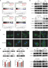
THEMIS2 promotes epithelial-mesenchymal transition and regulates F-actin assemble of ovarian cancer cells. (A) Gene set enrichment analysis (GSEA) plot based on the gene expression profililes of THEMIS2-high group compared with THEMIS2-low group using the TCGA and GSE9891 datasets. NES, normalized enrichment score. (B) The level of EMT markers in sh-NC vs. sh-THEMIS2 and vector vs. THEMIS2-OE Hey (top) and OVCAR8 (bottom) cells. (C) Visualization of F-actin by phalloidin staining in sh-THEMIS2 (left) or THEMIS2-OE (right) OC cells. Red arrows indicate lamellipodia and filopodia, while white arrows indicate impaired lamellipodia and filopodia formation. (D) THEMIS2 knockdown decreased the F-actin as in pellet (P) while increased the G-actin as in supernatant (S) in OC cells. Shown are representative immunoblot images (top) and summary data (bottom). (E) The level of cofilin and p-cofilin in Hey (left) and OVCAR8 (right) cells with THEMIS2 silencing or THEMIS2 overexpressing
Fig. 4
Fig. 4
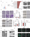
THEMIS2 enhances migrative and invasive ability of ovarian cancer cells via Rap1 signaling. (A) Volcano plot showed fold changes (x-axis) and corresponding p values (log10, y-axis) of genes in RNAseq data between control and THEMIS2 knockdown samples (Student’s t-test). (B) The KEGG pathways enriched in genes with THEMIS2 silencing. (C) Pull-down assays for active Rap1 in THEMIS2- knockdown or overexpression OC cells. (D) Transwell assays. Left: representative micrographs of the crystal violet-stained migrated THEMIS2-OE Hey and OVCAR8 cells treated with Rap1 inhibitor GGTI298. (E-F) Wound healing assays showed that migrative ability of cells was impended by GGTI298 in THEMIS2-OE OC cells. (G) Western blots of protein lysate of cultured Hey and OVCAR8 cells treated with GGTI298 or vehicle. (H) Representative photographs showing impaired cytoskeletal assembly in OC cells treated with GGTI298 or vehicle
Fig. 5
Fig. 5
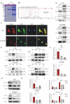
THEMIS2 regulates Rap1 signaling via maintaining combination between DOCK4 and CRKII. (A) Representative Coomassie blue staining images of anti-THEMIS2 or normal IgG immunoprecipitated proteins from Hey cells. (B) The result of IP-MS demonstrated that DOCK4 was identified to be a putative protein binding to THEMIS2. (C) Immunofluorescence assay was used to confirm THEMIS2’s colocalization with DOCK4 in Hey and OVCAR8 cells. (D) Co‐immunoprecipitation analysis was performed to detect the interaction of THEMIS2 with DOCK4. (E) Co-immunoprecipitation of DOCK4 and CRKII in OC cells with or without THEMIS2 silencing. (F-G) Validation of knockdown efficiency of DOCK4 in Hey and OVCAR8 cells through qRT-PCR and Western blot. The level of active Rap1-GTP was attenuated by si-DOCK4. (H-I) Trans-well and wound healing assays showed that knockdown of DOCK4 impaired the invasive and migrative ability of Hey and OVCAR8 cells
Fig. 6
Fig. 6
THEMIS2 mediates DOCK4-CRKII complex formation by recruiting TBK1 to phosphorylate DOCK4 at Ser1787. (A) DOCK4 phosphorylation is observed when endogenous DOCK4 is immunoprecipitated from Hey and OVCAR8 cells and analyzed by phospho-serine/threonine antibody. (B) The result of IP-MS demonstrated that TBK1 was identified to be a putative protein binding to THEMIS2. (C) Co-immunoprecipitation analysis was performed to detect the interaction of THEMIS2 with TBK1. (D) Co-immunoprecipitation analysis of DOCK4-TBK1 interaction in Hey and OVCAR8 cells following THEMIS2 knockdown or overexpression. (E) Treatment with TBK1 inhibitors GSK8612 inhibits phosphorylation of DOCK4. (F) Co-immunoprecipitation of DOCK4 and CRKII in OC cells with or without GSK8612 treatment. (G) The PhosphositePlus database indicated potential phosphorylation and ubiquitylation in the DOCK4 protein. (H) The prediction result of DOCK4 phosphorylation sites using the GPS 6.0 algorithm. (I) GSK8612 significantly inhibits serine/threonine phosphorylation of DOCK4WT but shows no observable inhibition of DOCK4S1787A serine/threonine phosphorylation in OC cells. (J) Activation of Rap1 following transient transfection of DOCK4WT, DOCK4S1787A and DOCK4S1787E into OC cells. (K) Co-immunoprecipitation of DOCK4 and CRKII in OC cells following transient transfection of DOCK4WT, DOCK4S1787A and DOCK4S1787E
Fig. 7
Fig. 7
Impact of Rap1 inhibition in an in vivo peritoneal metastasis model. (A) Overview of the animal experiment. (B) Representative bioluminescence photographs of mice after intraperitoneal inoculation of Hey-Luc cells with either over- or normal expression of THEMIS2 and treated with vehicle or GGTI298 (1.16 mg/kg). (C) Statistics for the total bioluminescence of the intraperitoneal xenograft OC mouse models (n = 5). (D) Body weight was measured weekly. (E) Representative views of the metastases in the peritoneal cavity are shown. Tumors were located on the intestine and liver. (F) Number of mice with intestinal, liver and diaphragmatic metastases. (G) The metastatic colonization was identified by hematoxylin and eosin staining. Arrows indicate tumors. (H) IHC staining for THEMIS2, E-cadherin, Vimentin, MMP2, and MMP9 of tumor tissues in each group
Fig. 8
Fig. 8
A model depicting THEMIS2-mediated promotion of OC metastasis. Increased of THEMIS2 in ovarian cancer promotes metastasis of this malignant tumor. The aberrant overexpression of THEMIS2 orchestrates metastatic progression in OC cells by recruiting TBK1 to phosphorylate DOCK4. This phosphorylation facilitates CRKII binding and subsequent Rap1 activation, which collectively drive EMT and F-actin polymerization

Similar articles

References

    1. F. Bray, M. Laversanne, H. Sung, J. Ferlay, R.L. Siegel, I. Soerjomataram, A. Jemal, Global cancer statistics 2022: GLOBOCAN estimates of incidence and mortality worldwide for 36 cancers in 185 countries. CA Cancer J. Clin. 74, 229–263 (2024). 10.3322/caac.21834 - PubMed
    1. R.L. Siegel, K.D. Miller, N.S. Wagle, A. Jemal, Cancer statistics, 2023. CA Cancer J. Clin. 73, 17–48 (2023). 10.3322/caac.21763 - PubMed
    1. D. Dhaliwal, T.G. Shepherd, Molecular and cellular mechanisms controlling integrin-mediated cell adhesion and tumor progression in ovarian cancer metastasis: a review. Clin. Exp. Metastasis. 39, 291–301 (2022). 10.1007/s10585-021-10136-5 - PMC - PubMed
    1. K.D. Miller, L. Nogueira, T. Devasia, A.B. Mariotto, K.R. Yabroff, A. Jemal, J. Kramer, R.L. Siegel, Cancer treatment and survivorship statistics, 2022. CA Cancer J. Clin. 72, 409–436 (2022). 10.3322/caac.21731 - PubMed
    1. J. Yang, P. Antin, G. Berx, C. Blanpain, T. Brabletz, M. Bronner, K. Campbell, A. Cano, J. Casanova, G. Christofori et al., Guidelines and definitions for research on epithelial-mesenchymal transition. Nat. Rev. Mol. Cell. Biol. 21, 341–352 (2020). 10.1038/s41580-020-0237-9 - PMC - PubMed

Substances