Review
. 2025 Mar 23;17(7):1077.
doi: 10.3390/cancers17071077.

The Role of Perirenal Adipose Tissue in Carcinogenesis-From Molecular Mechanism to Therapeutic Perspectives

Affiliations
Review

The Role of Perirenal Adipose Tissue in Carcinogenesis-From Molecular Mechanism to Therapeutic Perspectives

Adriana Grigoraș et al. Cancers (Basel). .

Abstract

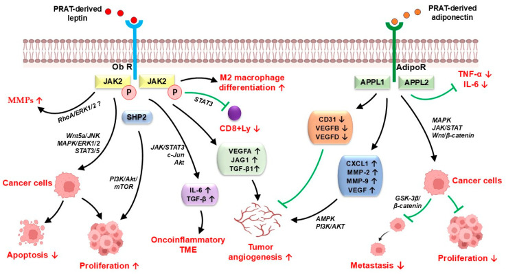
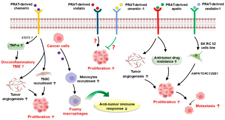
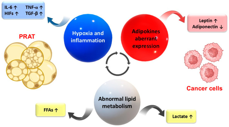
Perirenal adipose tissue (PRAT) exhibits particular morphological features, with its activity being mainly related to thermogenesis. However, an expanded PRAT area seems to play a significant role in cardiovascular diseases, diabetes mellitus, and chronic kidney disease pathogenesis. Numerous studies have demonstrated that PRAT may support cancer progression and invasion, mainly in obese patients. The mechanism underlying these processes is of dysregulation of PRAT's secretion of adipokines and pro-inflammatory cytokines, such as leptin, adiponectin, chemerin, apelin, omentin-1, vistatin, nesfatin-1, and other pro-inflammatory cytokines, modulated by tumor cells. Cancer cells may also induce a metabolic reprogramming of perirenal adipocytes, leading to increased lipids and lactate transfer to the tumor microenvironment, contributing to cancer growth in a hypoxic milieu. In addition, the PRAT browning process has been specifically detected in renal cell carcinoma (RCC), being characterized by upregulated expression of brown/beige adipocytes markers (UCP1, PPAR-ɣ, c/EBPα, and PGC1α) and downregulated white fat cells markers, such as LEPTIN, SHOX2, HOXC8, and HOXC9. Considering its multifaceted role in cancer, modulation of PRAT's role in tumor progression may open new directions for oncologic therapy improvement. Considering the increasing evidence of the relationship between PRAT and tumor cells, our review aims to provide a comprehensive analysis of the perirenal adipocytes' impact on tumor progression and metastasis.

Keywords: adipokines; cancer; cancer therapy; metastasis; perirenal adipose tissue; predictive factor; prognosis.


Conflict of interest statement

The authors declare no conflicts of interest.

Figures

Figure 1
Figure 1
Potential mechanisms of PRAT-derived leptin and adiponectin in cancer progression and metastasis. PRAT-derived leptin binds to its receptor (Ob-R), promoting tumor growth and metastasis by JAK/STAT3, PI3K/Akt/mTOR, Wnt5a/JNK, and MAPK/ERK1/2 pathways, modulates the anti-tumor immune response by M2 macrophage differentiation, and is involved in extracellular matrix degradation via RhoA/ERK1/2 signaling stimulation. PRAT-derived adiponectin binds to its receptor (AdipoR), leading to the recruitment of APPL1 and APPL2, followed by the activation of several signaling pathways associated with anti-tumor effects and reduction of the tumor microenvironment inflammatory features by decreased expression of TNF-α and IL-6, but it also promotes tumor angiogenesis via AMPK and PI3K/AKT pathway activation. Adipo-R—adiponectin receptor; CD31—cluster differentiation 31; CXCL1—chemokine (C-X-C motif) ligand 1; IL-6—Interleukin 6; MMP—matrix metalloproteinase; Ob-R—leptin receptor; PRAT—perirenal adipose tissue; TME—tumor microenvironment; TNF-α—tumor necrosis factor α; VEGFA—vascular endothelial growth factor A; VEGFB—vascular endothelial growth factor B; ↑—increase; ↓—decrease; →—activation; ꓕ—inhibition.
Figure 2
Figure 2
Potential mechanisms of PRAT-derived chemerin, visfatin, omentin-1, apelin and nesfatin-1 in cancer progression and metastasis. PRAT-derived chemerin promotes tumor angiogenesis, tumor-supporting stromal cell recruitment, contributes to the tumor’s pro-inflammatory features by TNF-α expression increase via STAT3 pathway activation, and may modulate the anti-tumor immune response by monocyte recruitment and their change into foamy macrophages. PRAT-derived visfatin, omentin-1, and apelin support cancer cell proliferation, while PRAT-derived nesfatin-1 may promote tumor cell metastasis by AMPK/TORC1/ZEB1 pathway activation. PRAT—perirenal adipose tissue; TNF-α—tumor necrosis factor α; TSSC—tumor-supporting stromal cells; ↑—increase; ↓—decrease; →—activation; ꓕ—inhibition.
Figure 3
Figure 3
Potential mechanisms linking cancer to PRAT dysfunction in the tumor microenvironment. Aberrant adipokine expression and abnormal lipid metabolism, along with the increased hypoxic and inflammatory features of the tumor microenvironment, are the main mechanisms of tumor cell–PRAT interrelationship. FAAs—free fatty acids; HIFs—hypoxia-inducible factors; IL-6—Interleukin 6; PRAT—perirenal adipose tissue; TGF-β—transforming growth factor-β; TNF-α—tumor necrosis factor α; ↑—increase; ↓—decrease.

Similar articles

References

    1. Fang Y., Xu Y., Yang Y., Liu C., Zhao D., Ke J. The relationship between perirenal fat thickness and reduced glomerular filtration rate in patients with type 2 diabetes. J. Diabetes Res. 2020;2020:6076145. doi: 10.1155/2020/6076145. - DOI - PMC - PubMed
    1. Kim J.W., Kim J.H., Lee Y.J. The role of adipokines in tumor progression and its association with obesity. Biomedicines. 2024;12:97. doi: 10.3390/biomedicines12010097. - DOI - PMC - PubMed
    1. Grigoras A., Balan R.A., Caruntu I.-D., Giusca S.E., Lozneanu L., Avadanei R.E., Rusu A., Riscanu L.A., Amalinei C. Perirenal adipose tissue—Current knowledge and future opportunities. J. Clin. Med. 2021;10:1291. doi: 10.3390/jcm10061291. - DOI - PMC - PubMed
    1. Jespersen N.Z., Feizi A., Andersen E.S., Heywood S., Hattel H.B., Daugaard S., Peijs L., Bagi P., Feldt-Rasmussen B., Schultz H., et al. Heterogeneity in the perirenal region of humans suggests presence of dormant brown adipose tissue that contains brown fat precursor cells. Mol. Metab. 2019;24:30–43. doi: 10.1016/j.molmet.2019.03.005. - DOI - PMC - PubMed
    1. Fazeli S.A., Nourollahi S., Alirezaei A., Mirhashemi S., Davarian A., Hosseini I. Perirenal adipose tissue: Clinical implication and therapeutic interventions. Indian J. Nephrol. 2024;34:573–582. doi: 10.25259/ijn_532_23. - DOI - PMC - PubMed

LinkOut - more resources