Review
. 2025 Mar 30;17(7):1167.
doi: 10.3390/cancers17071167.

Exosome-Mediated Cellular Communication in the Tumor Microenvironment Imparts Drug Resistance in Breast Cancer

Affiliations
Review

Exosome-Mediated Cellular Communication in the Tumor Microenvironment Imparts Drug Resistance in Breast Cancer

RamaRao Malla et al. Cancers (Basel). .

Abstract

Globally, breast cancer (BC) is the leading cause of cancer-related death for women. BC is characterized by heterogeneity, aggressive behavior, and high metastatic potential. Chemotherapy, administered as monotherapy or adjuvant therapy, remains a cornerstone of treatment; however, acquired drug resistance is a significant clinical challenge. Deciphering mechanisms of drug resistance will be central to developing more efficient treatment options and improving patient outcomes. The current review examines the multifaceted nature of exosomes in conferring drug resistance in BC through complex communication networks within the tumor microenvironment. We further explore recent advances in understanding how exosomes contribute to resistance against established chemotherapeutic agents such as tamoxifen, paclitaxel, doxorubicin, platinum-based drugs, trastuzumab, and newer immunotherapies, such as immune checkpoint inhibitors. Moreover, we discuss existing systematic approaches to investigating the exosome-drug resistance relationship in BC. Finally, we explore promising therapeutic approaches to overcome exosome-dependent drug resistance in BC, highlighting potential avenues for improved treatment efficacy. Investigating the distinct functions and cargo of exosomes offers potential for developing innovative approaches to overcoming treatment resistance.

Keywords: breast cancer; drug resistance; drug sensitization; exosomes; tumor microenvironment.


Conflict of interest statement

The authors declare no conflicts of interest.

Figures
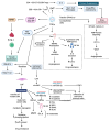
Figure 1
Figure 1
Interplay of exosomes and cellular communication in the BC TME. Tumor cell-derived exosomes (TDEs) initiate pre-metastatic niche formation in the lungs by transferring caveolin-1. This transfer induces the expression of premetastatic niche-associated genes in lung epithelial cells, stimulates tenascin-C secretion, and promotes extracellular matrix (ECM) deposition. Concurrently, TDEs facilitate macrophage (MØ) polarization to M2-type macrophages, which secrete VEGF-A to promote angiogenesis in lung tissues. Additionally, TDEs reprogram tumor cell metabolism by transferring miR-122, increasing nutrient availability to support metastasis. TDEs promote a pro-tumoral M2-like phenotype by activating NF-κB signaling, which leads to IL-6 secretion, which in turn activates toll-like receptor 2 (TLR2)-mediated pro-inflammatory response within the TME. TDEs suppress the anti-tumor immune response by reprogramming cytotoxic T cells (TC cells) via transfer of cirmiR-20a-5p. This transfer results in reduced expression of nuclear protein ataxia-telangiectasia (NPAT), which causes TC cell dysfunction by enhancing PD-1 expression and reducing glycolysis via disrupting AKT-mTOR signaling. TDEs also suppress T cells via reducing CD3+HLA-DR+ T cells, increasing CD3+PD-L1 T cells, and promoting IL-10 secretion from CD4+CD127-CD25hi Tregs. Drug-resistant cancer cells induce drug resistance phenotypes in sensitive cells by transferring exosomes that induce epithelial-to-mesenchymal transition (EMT), activate stem cell-like programs, modulate drug efflux mechanisms, and activate survival signals. TDEs facilitate angiogenesis by transferring ephrin type-A receptor 2 (EPHA2) to endothelial cells, thereby increasing vesicular endothelial growth factor (VEGF) expression through AMP-activated protein kinase (AMPK) signaling and HIF-1α activation. Cancer-associated adipocytes (CAAs) induce drug resistance and metastasis by transferring circular RNA cysteine-rich transmembrane bone morphogenetic protein regulator 1 (circCRIM1). This results in inhibition of miR-503-5p, which in turn activates glycosylation hydrolase, which destabilizes fructose-1,6-bisphosphatase 1 (FBP1). Both changes further promote immune cell infiltration. TDEs reprogram normal fibroblasts into cancer-associated fibroblasts (CAFs) by transferring small nucleolar RNA host gene 14 (SNHG14), which upregulates family with sequence similarity 171, member A1 (FAM171A1) expression through early B-cell factor 1 (EBF1). CAFs promote tumor progression by releasing exosomes. These exosomes transfer miR-500a-5p, which directly targets ubiquitin-specific peptidase 26 (USP26), leading to downregulation of E-cadherin and upregulation of N-cadherin, fibronectin 1 (FN1), zinc-finger E-box binding homeobox 1 (ZEB1), Snail, and Slug. Exosomes from CAFs transfer mutant mitochondrial DNA, which triggers quiescent cancer cells to exit dormancy, reprogramming their metabolism to abolish oxidative phosphorylation and increase self-renewal. CAFs also promote invasion by transferring autophagy-associated G-protein-coupled receptor 64 (GPR64), which upregulates matrix metalloproteinase 9 (MMP9) and interleukin-8 (IL-8) in recipient BC cells by stimulating non-canonical NF-κB signaling.
Figure 2
Figure 2
Exosome-mediated tamoxifen resistance in BC. Exosomes impart tamoxifen (TMX) resistance in several ways. By circular RNA encoding ubiquitin-conjugating enzyme E2 D2 (circUBE2D2) transfer, they promote BC metastasis. By mi-R-9-5p, miR205 transfer, and urothelial carcinoma associated 1 (UCA1) enriched exosomes, TMX resistance is induced by inhibiting apoptosis. Exosomes also maintain the cancer-associated fibroblast (CAF) phenotype by transferring CD63 via signal transducer and activator of transcription 3 (STAT3) signaling and by serine/arginine-rich splicing factor 1 (SFRS1) through miR-22 packing into them. Exosomal heat shock protein family H (Hsp110) member 1 (HSPH1) induces TMX resistance in MCF-7 cells. ETF1/miR205 induces TMX resistance by activating AKT, which inhibits caspase-3, reduces apoptosis, and enhances drug resistance.
Figure 3
Figure 3
Exosome-mediated DOX resistance in BC. DOX induces toxicity in cancer cells by inhibiting cell proliferation, inducing DNA double-strand breaks (DBS), and inhibiting topoisomerase II-induced cell death by inducing ROS generation. Cancer cells develop resistance against DOX by increasing drug efflux through overexpressing ABC transporters and overexpressing miR181b-5p, miR582-3p, miR-221-3p, miR-222, miR 155, and miR 34a-5p, which promote apoptosis inhibition, cell proliferation, tumor promotion, M2 macrophage polarization, and stem cell induction. These miRs are lodged into exosomes and transported to DOX sensitive cells to induce resistance by multiple signaling mechanisms, as depicted in the figure. DOX inhibits cell proliferation by activating the cAMP responsive element binding protein-like 1 (CREBL1)/proliferation potential protein (P2P) pathway via enhancing synthesis of ceramide. Ring finger-11 (RFN-11) promotes ubiquitination and degradation of proapoptotic proteins; phosphatase and tensin homolog deleted on chromosome 10 (PTEN-10)/forkhead box O (FOXO) activates M2 macrophage polarization.
Figure 4
Figure 4
Exosome-mediated paclitaxel (PTX) resistance in BC: PTX induces toxicity in cancer cells by inhibiting tubulin to disrupt microtubule stability and affecting the process of cell division. It also activates procaspases and proapoptotic proteins, leading to cell death. Cancer cells develop resistance against PTX, increasing drug efflux through overexpressing ABC transporters (p-glycoprotein, multidrug resistance protein 1 and breast cancer resistance protein) and overexpressing miR217, miR378a-3p, miR378d, miR 155, annexin A6 (Anx6), heat shock protein 90 (HSP90), and survivin, which promote drug efflux, metastasis, EMT transition, stem cell induction, and tumor progression. These miR molecules and Anx6, HSP90, and survivin are lodged into exosome cargo and transported to PTX-sensitive cells to transfer resistance via multiple signaling mechanisms, as depicted in the figure. Exosomes mediate drug resistance by inhabiting apoptosis by activating Taxol resistance–associated gene-3 (TRAG-3)/chondrosarcoma-associated gene 2 (CSAG2).
Figure 5
Figure 5
Exosome-mediated platinum drug resistance in breast cancer. Oxaplatin, carboplatin, cisplatin, lobaplatin, and nedaplatin are different platinum drugs used to treat different cancers. Tumor cell exosomes upregulate p-glycoprotein, which pumps the drug out of the cell; miR-423-5p alters the TME; exosomes released by Snail-induced EMT inhibit the activity of PTEN and BRCC3, thus downregulating the activity of NLRP3; the exosomes derived from SOD1 fibroblasts release miR-3960; this miRNA targets the BRSK2-induced phosphorylation of PIMREG by S16, thus inhibiting or decreasing the activity of the NF-Kβ signaling pathway—all these mechanisms by exosomes lead to cisplatin resistance. Note: Red bar denotes inhibition, downward red arrow denotes down-regulation, upward green arrow denotes upregulation.
Figure 6
Figure 6
Exosome-mediated trastuzumab resistance in breast cancer. Exosomal Linc00969 induces HER-2 gene upregulation and promotes TRA resistance by triggering autophagy. Oncogenic circHIPK3 is regulated directly by miR-582-3p, which regulates the expression of RNF11, which induces drug resistance by selective degradation of proapoptotic proteins. Exosomes containing neuropeptide neuromedin U (NmU) enhance the release of TGF-β and PD-L1. TGF-β induces TRA resistance, and PD-L1 induces immune suppression in BC cells.

References

    1. Ugai T., Sasamoto N., Lee H.Y., Ando M., Song M., Tamimi R.M., Kawachi I., Campbell P.T., Giovannucci E.L., Weiderpass E., et al. Is early-onset cancer an emerging global epidemic? Current evidence and future implications. Nat. Rev. Clin. Oncol. 2022;19:656–673. doi: 10.1038/s41571-022-00672-8. - DOI - PMC - PubMed
    1. Lei S., Zheng R., Zhang S., Wang S., Chen R., Sun K., Zeng H., Zhou J., Wei W. Global patterns of breast cancer incidence and mortality: A population-based cancer registry data analysis from 2000 to 2020. Cancer Commun. 2021;41:1183–1194. doi: 10.1002/cac2.12207. - DOI - PMC - PubMed
    1. Liang Y., Zhang H., Song X., Yang Q. Metastatic heterogeneity of breast cancer: Molecular mechanism and potential therapeutic targets. Semin. Cancer Biol. 2020;60:14–27. doi: 10.1016/j.semcancer.2019.08.012. - DOI - PubMed
    1. Lee J., Park Y.H. Trastuzumab deruxtecan for HER2+ advanced breast cancer. Future Oncol. 2022;18:7–19. doi: 10.2217/fon-2021-0550. - DOI - PubMed
    1. Lev S. Targeted therapy and drug resistance in triple-negative breast cancer: The EGFR axis. Biochem. Soc. Trans. 2020;48:657–665. doi: 10.1042/BST20191055. - DOI - PubMed

LinkOut - more resources