. 2025 Jun;12(21):e2414792.
doi: 10.1002/advs.202414792. Epub 2025 Apr 14.

Dimethyl Itaconate Alleviates Escherichia coli-Induced Endometritis Through the Guanosine-CXCL14 Axis via Increasing the Abundance of norank_f_Muribaculaceae

Affiliations

Dimethyl Itaconate Alleviates Escherichia coli-Induced Endometritis Through the Guanosine-CXCL14 Axis via Increasing the Abundance of norank_f_Muribaculaceae

Yuhang He et al. Adv Sci (Weinh). 2025 Jun.

Abstract

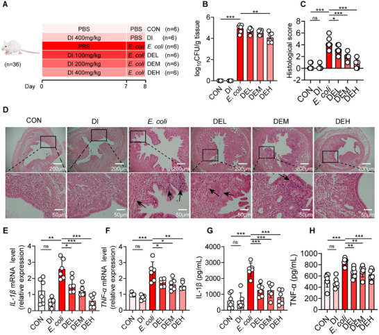
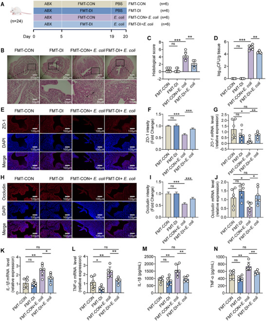
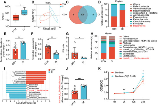
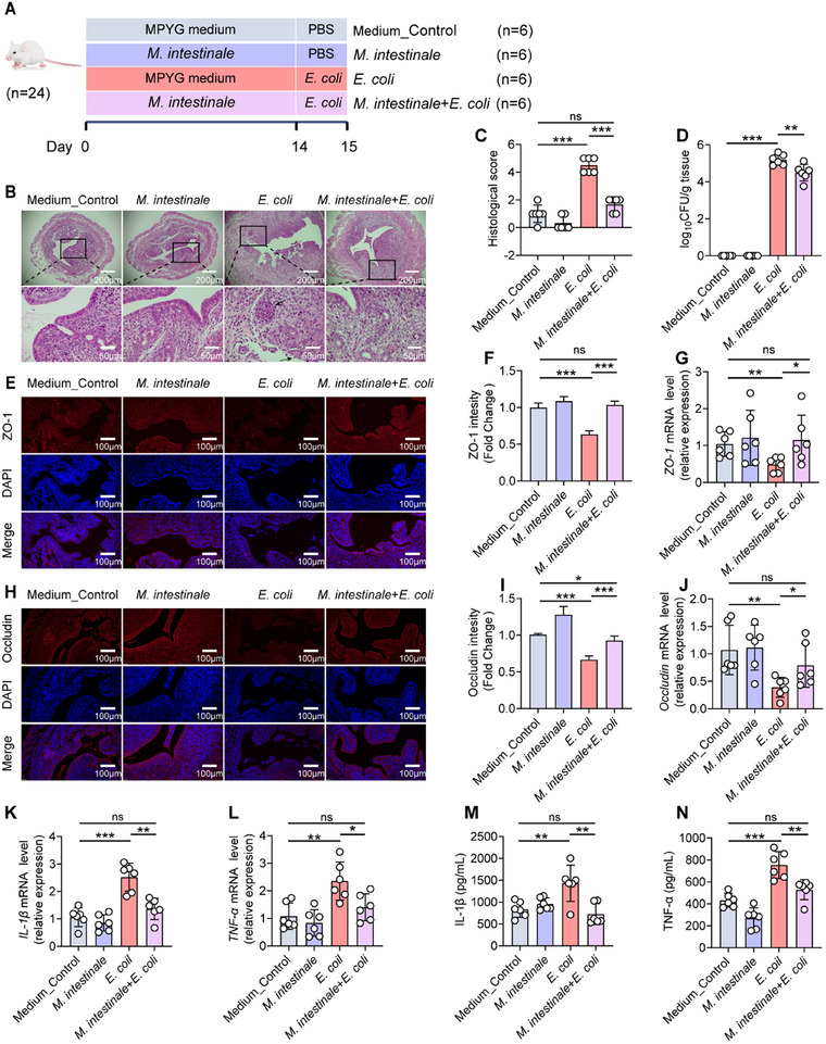
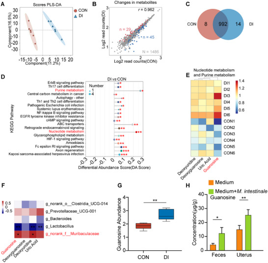
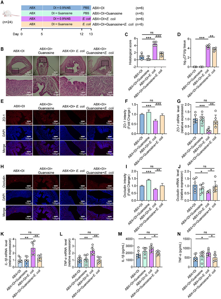
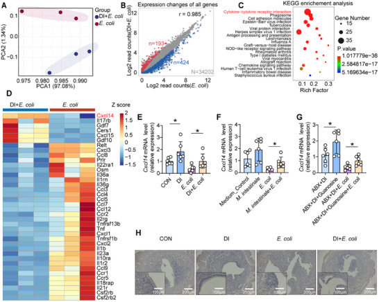
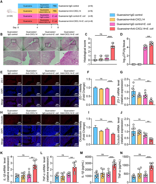
Endometritis, a prevalent reproductive system disease with high incidence, leads to reproductive dysfunction in humans and animals, causing huge economic losses. Dimethyl itaconate (DI) has been demonstrated to exert protective effects in multiple inflammatory diseases. Nevertheless, the efficacy of DI in preventing endometritis and the role played by the gut microbiota remain unknown. In this study, it is found that DI ameliorated Escherichia coli (E. coli) induced endometritis in mice. The protective effect is abolished by antibiotic-induced depletion of the gut microbiota, and fecal microbiota transplantation (FMT) from DI-treated mice to recipient mice ameliorated E. coli-induced endometritis. Integrative multiomics reveals that DI promotes the multiplication of norank_f_Muribaculaceae in vivo, and supplementation of Muribaculum intestinale (DSM 28989), which belongs to the norank_f_Muribaculaceae genus, upregulates the level of guanosine in the uterus. Mechanistically, the protective effect of guanosine in endometritis is mediated by activating the expression of CXCL14 in uterine epithelial cells. Moreover, the antibody-neutralizing experiment of CXCL14 eliminated this protective effect. In conclusion, this study elucidates the significant role of the gut microbiota and its metabolites in the protection of DI against endometritis, and provides new evidence for the regulation of distal organ by the gut microbiota.

Keywords: dimethyl itaconate; endometritis; gut microbiota; gut‐uterus axis.


Conflict of interest statement

The authors declare no conflict of interest.

Figures

Figure 1
Figure 1
Dimethyl itaconate (DI) dose‐dependently attenuated E. coli‐induced endometritis in mice. A–H) The mice were divided into six groups and treat them with different concentrations of DI (0, 100, 200, or 400 mg kg−1) for 7 days. Four groups of different concentrations of DI were followed by uterine injection of 10⁸ CFU of E. coli to induce endometritis. Six groups were generated: CON, DI (400 mg kg−1), E. coli, DEL (100 mg kg−1 DI+E. coli), DEM (200 mg kg−1 DI+E. coli) and DEH (400 mg kg−1 DI+E. coli) groups. A) Study design of in vivo mouse experiment. B) The concentration of E. coli in the uterus was determined by plate coating (n = 6). C) Histological scores in different treatment groups were performed (n = 6). D) Representative images of the H&E‐stained uterus sections of indicated groups. The black arrow indicates endometrial injury. E) The mRNA expression of IL‐1β in uterine tissue. F) The mRNA expression of TNF‐α in uterine tissue. G) IL‐1β levels in uterine tissue homogenate by ELISA. H) TNF‐α levels in uterine tissue homogenate by ELISA. Data represent means ± SD; * p < 0.05; ** p < 0.01; *** p < 0.001; by unpaired Student's t test. The data shown are representative of three independent experiments.
Figure 2
Figure 2
Fecal microbiota transplantation (FMT) from oral administration of dimethyl itaconate (DI) group can alleviate E. coli‐induced endometritis in mice. A–N) Mice were administered antibiotics for 5 days to remove gut microbiota, and then colonized with gut microbiota from CON and DI groups for 14 days. Afterwards, the mice were followed by uterine injection of 10⁸ CFU of E. coli to induce endometritis (n = 6). A) FMT experimental design. B) Representative images of the H&E‐stained uterus sections of indicated groups. The black arrow indicates endometrial injury. C) Histological scores in different treatment groups were performed (n = 6). D) The concentration of E. coli in the uterus was determined by plate coating. E,F) Uterine sections were immunofluorescent staining with ZO‐1, and the nuclei were visualized by DAPI staining. G) The mRNA expression of ZO‐1 in uterine tissue. H–I) Uterine sections were immunofluorescent staining with Occludin, and the nuclei were visualized by DAPI staining. J) The mRNA expression of Occludin in uterine tissue. K) The mRNA expression of IL‐1β in uterine tissue. L) The mRNA expression of TNF‐α in uterine tissue. M) IL‐1β levels in uterine tissue homogenate by ELISA. N) TNF‐α levels in uterine tissue homogenate by ELISA. Data represent means ± SD; * p < 0.05; ** p < 0.01; *** p < 0.001; by unpaired Student's t test. The data shown are representative of three independent experiments.
Figure 3
Figure 3
Oral administration of dimethyl itaconate (DI) regulated the composition gut microbiota in mice. A–J) Mice were given DI orally or distilled water for 7 days. Feces from the CON group and DI group mice were collected for 16S rRNA sequencing (n = 6). A) The α‐diversity analysis of gut microbes reflected by Chao1 indice. B) Scatter plots of weighted PCoA for the microbial composition showed the differences in gut microbial structure between the CON and DI groups. C) Venn diagram of the amplicon sequence variants (ASVs) in the CON and DI groups. D) Relative abundance of gut microbiota at the phylum level from different treatment groups. E) The abundance of Bacteroidetes between the CON and DI groups. F) The abundance of Firmicutes between the CON and DI groups. G) The ratio of F/B of CON and DI groups. H) Relative abundance of gut microbiota at the genus level from different treatment groups. I) LEfSe showed different bacterial taxa that were enriched in different groups (log10 LDA score >4). J) The abundance of the genera norank_f_Muribaculaceae between the CON and DI groups. K) The growth curve of the M. intestinale cultured with DI (0.5 × 10⁻³ m). Data represent means ± SD; * p < 0.05; ** p < 0.01; *** p < 0.001; compared with the CON group.
Figure 4
Figure 4
Administration of M. intestinale alleviated E. coli‐induced endometritis in mice. A–N) Mice were gavaged with M. intestinale daily for 2 weeks. Equivalent sterile MPYG medium was used as vehicle control. Afterwards, mice were followed by uterine injection of 10⁸ CFU of E. coli to induce endometritis. (A) M. intestinale administration experimental design. B) Representative images of the H&E‐stained uterus sections of indicated groups. The black arrow indicates endometrial injury. C) Histological scores in different treatment groups were performed (n = 6). D) The concentration of E. coli in the uterus was determined by plate coating. E,F) Uterine sections were immunofluorescent staining with ZO‐1, and the nuclei were visualized by DAPI staining. G) The mRNA expression of ZO‐1 in uterine tissue. H–I) Uterine sections were immunofluorescent staining with Occludin, and the nuclei were visualized by DAPI staining. J) The mRNA expression of Occludin in uterine tissue. K) The mRNA expression of IL‐1β in uterine tissue. L) The mRNA expression of TNF‐α in uterine tissue. M) IL‐1β levels in uterine tissue homogenate by ELISA. N) TNF‐α levels in uterine tissue homogenate by ELISA. Data represent means ± SD; * p < 0.05; ** p < 0.01; *** p < 0.001; by unpaired Student's t test. The data shown are representative of three independent experiments.
Figure 5
Figure 5
Dimethyl itaconate (DI) altered the metabolic profiling and enriched microbiota‐derived purine metabolites–guanosine. A–G) Mice were orally administered distilled water or DI for 7 days. Feces from the CON group and DI group mice were collected for untargeted metabolomics (n = 6). A) Partial least‐squares discriminant analysis (PLS‐DA) was performed on the metabonomics profiles in fecal samples from the DI and CON groups. B) Volcano plot of differentially expressed genes. C) Venn diagrams. D) KEGG pathway difference abundance score chart. E) Heat map of significantly altered metabolites in feces. F) Correlation analysis between fecal metabolites exhibiting significant changes and differential gut microbiota. G) Distribution box plot of metabolite guanosine. H) The abundance of guanosine in the feces and uterus of mice from M. intestinale administration experiment (3–4 biological replicates for each group). The red color denotes a positive correlation, while blue color denotes a negative correlation. The intensity of the color is proportional to the strength of Spearman correlation. Data represent means ± SD; * p < 0.05; ** p < 0.01; *** p < 0.001; by unpaired Student's t test.
Figure 6
Figure 6
Dimethyl itaconate (DI), rather than directly exerting anti‐inflammatory effects, mediates protective effects against E. coli‐induced endometritis through the metabolite guanosine produced by the gut microbiota. A–N) Mice were pretreated with ABX for 5 days to deplete the gut microbiota. Subsequently, DI was administered orally. Meanwhile, guanosine dissolved in 0.9% normal saline (0.9% NS) at a dose of 8 mg kg−1 was given by intraperitoneal injection for 7 days, followed by uterine injection with 108 CFU of E. coli to induce endometritis (n = 6). A) Guanosine supplementation experimental design. B) Representative images of the H&E‐stained uterus sections of indicated groups. The black arrow indicates endometrial injury. C) Histological scores in different treatment groups were performed (n = 6). D) The concentration of E. coli in the uterus was determined by plate coating. E,F) Uterine sections were immunofluorescent staining with ZO‐1, and the nuclei were visualized by DAPI staining. G) The mRNA expression of ZO‐1 in uterine tissue. H–I) Uterine sections were immunofluorescent staining with Occludin, and the nuclei were visualized by DAPI staining. J) The mRNA expression of Occludin in uterine tissue. K) The mRNA expression of IL‐1β in uterine tissue. L) The mRNA expression of TNF‐α in uterine tissue. M) IL‐1β levels in uterine tissue homogenate by ELISA. N) TNF‐α levels in uterine tissue homogenate by ELISA. Data represent means ± SD; * p < 0.05; ** p < 0.01; *** p < 0.001; by unpaired Student's t test. The data shown are representative of three independent experiments.
Figure 7
Figure 7
Dimethyl itaconate (DI) alters gene expression in the mouse uterus. A–H) Mice were orally administered distilled water or DI for 7 days, followed by induction of endometritis using E. coli (n = 3). A) Principal component analysis (PCA) of the transcriptome of mouse uterine tissue. B) Dot plot of differentially expressed genes in the uterus after oral administration of DI. C) Enrichment analysis of the most significantly altered pathways in the KEGG pathway. D) Heatmap analysis of the enriched differential genes in the cytokine‐cytokine receptor interaction pathway. E–G) Relative expression changes of CXCL14 in the previous experiments by RT‐qPCR. H) Representative images of immunohistochemistry staining with CXCL14. Data are mean ± SD. * p < 0.05, ** p < 0.01, and *** p < 0.001. Statistical analysis was performed using Student's t test.
Figure 8
Figure 8
The protective effect of guanosine against E. coli‐induced endometritis disappears when systemic neutralization of CXCL14 is performed. A–N) Mice received intraperitoneal injections of guanosine for seven consecutive days, followed by intraperitoneal injection of Anti‐CXCL14 for the last 3 days to neutralize endogenous CXCL14. Subsequently, mice were followed by uterine injection of 10⁸ CFU of E. coli to induce endometritis. A) Experimental design for CXCL14 inhibition. B) Representative images of the H&E‐stained uterus sections of indicated groups. The black arrow indicates endometrial injury. C) Histological scores in different treatment groups were performed (n = 6). D) The concentration of E. coli in the uterus was determined by plate coating. E,F) Uterine sections were immunofluorescent staining with ZO‐1, and the nuclei were visualized by DAPI staining. G) The mRNA expression of ZO‐1 in uterine tissue. H–I) Uterine sections were immunofluorescent staining with Occludin, and the nuclei were visualized by DAPI staining. J) The mRNA expression of Occludin in uterine tissue. K) The mRNA expression of IL‐1β in uterine tissue. L) The mRNA expression of TNF‐α in uterine tissue. M) IL‐1β levels in uterine tissue homogenate by ELISA. N) TNF‐α levels in uterine tissue homogenate by ELISA. Data represent means ± SD; * p < 0.05; ** p < 0.01; *** p < 0.001; by unpaired Student's t test. The data shown are representative of three independent experiments.

Similar articles

References

    1. Umar T., Yin B., Umer S., Ma X., Jiang K., Umar Z., Akhtar M., Shaukat A., Deng G., Inflammation 2021, 44, 1683. - PubMed
    1. Ravel J., Moreno I., Simón C., Am. J. Obstet. Gynecol. 2021, 224, 251. - PubMed
    1. Sheldon I. M., Noakes D. E., Rycroft A. N., Pfeiffer D. U., Dobson H., Reproduction 2002, 123, 837. - PubMed
    1. Dohmen M. J., Joop K., Sturk A., Bols P. E., Lohuis J. A., Theriogenology 2000, 54, 1019. - PubMed
    1. Raheel I., Hassan W. H., Salem S. S. R., Salam H. S. H., J. Adv. Vet. Anim. Res. 2020, 7, 442. - PMC - PubMed

LinkOut - more resources