. 2025 Mar 28:(217):10.3791/67364.
doi: 10.3791/67364.

Optogenetic Activation of Intrinsic Cardiac Autonomic Neurons in Excised Perfused Mouse Hearts

Affiliations

Optogenetic Activation of Intrinsic Cardiac Autonomic Neurons in Excised Perfused Mouse Hearts

Rebekah Russo et al. J Vis Exp. .

Abstract

A balance of cholinergic and catecholaminergic activation is necessary to maintain heart health. Interrogating the interaction between these pathways can be done using optogenetics through selective expression of channelrhodopsin-2 (ChR2) in cardiac autonomic neurons. Such cardiac applications of optogenetics allow for the study of the intrinsic release of neurotransmitters in a spatiotemporal manner. This method illustrates an ex vivo approach for specific optogenetic stimulation of cardiac neurons in perfused mouse hearts. Transgenic mice were bred to express ChR2 in either choline acetyltransferase (ChAT) or tyrosine hydroxylase (TH) neurons throughout the body. A micro-LED (465 nm) encased in a silicone elastomer was prepared for stimulating the neurons of the right atrium that innervate the sinoatrial node. The micro-LED was connected to a function generator set to pulse waves at 10 Hz with a 30 ms pulse width. Hearts with confirmed expression were excised and retrogradely perfused on a Langendorff system circulating Krebs-Henseleit solution. Electrocardiogram (ECG), temperature, and coronary flow rate were recorded using the LabChart software. Once the heart stabilized, the micro-LED was placed on the right atrium and tested for optimal heart rate response. An application of this approach combines the intrinsic release of cholinergic neurotransmitter (acetylcholine) during optogenetic activation of a ChAT-ChR2 mouse heart simultaneously with increasing exogenous catecholaminergic neurotransmitter (norepinephrine) added to the perfusate. The resulting changes in heart rate during the simultaneous cholinergic and catecholaminergic activation are presented. This method describes a valuable experimental approach for investigating the kinetics of sudden intrinsic autonomic neuron activation in perfused hearts and the interactions between cardiac cholinergic and catecholaminergic activity.


Conflict of interest statement

Disclosures

No conflicts of interest to declare.

Figures

Figure 1:
Figure 1:. Micro-LED construction.
Simplified overview of steps for constructing micro-LED light source. Two wires are soldered to the micro-LED and inserted into a 200 μL pipette tip, then superglued (Step 1). Silicone elastomer is mixed at a 10:1 ratio and placed in a vacuum chamber to remove bubbles (Step 2). The silicone elastomer is poured into a microcentrifuge tube, and the micro-LED pipette tip is inserted and allowed to cure overnight (Step 3). The insulated micro-LED is then removed from the tube, and excess silicone should be trimmed (Step 4).
Figure 2:
Figure 2:. Transgenic mouse breeding scheme.
A parent mouse with the lox-dependent ChR2 gene is crossbred with another mouse with a Cre promoter. A parent mouse with tyrosine hydroxylase (TH) Cre promoter will produce heterozygous offspring where 50% will express ChR2 in catecholaminergic cells. A parent mouse with choline acetyltransferase (ChAT) Cre promoter will produce homozygous offspring where 100% will express ChR2 in cholinergic cells. Expression is confirmed via genotyping. This figure has been modified with permission from.
Figure 3:
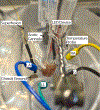
Figure 3:. Mouse heart bath configuration.
A cannulated mouse heart showing placement of LED device and ECG electrodes. A chassis ground is present to reduce noise from surrounding electronics. ECG needle electrodes are placed based on Einthoven’s triangle. Abbreviations: LA = left arm; RA = right arm; LL = left leg; G = ground.
Figure 4:
Figure 4:. Experimental setup. Diagram of perfusion system.
Arrows show the direction of perfusate. Superfusion perfusate is indicated by dashed lines and components are red outlined.
Figure 5:
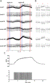
Figure 5:. Representative cholinergic photostimulation response.
(A) A 6 lead ECG during ChAT-ChR2 optogenetic activation. Solid blue lines indicate the micro-LED being turned on/off. Red dashed boxes indicate the time for (B) snippets. (B) Half-second snippets of ECG signal before (a), during (b), and after photostimulation (c). RR interval is shown for each section. (C) Heart rate (top) is shown along with pulse waves from a function generator (bottom). The heart rate starts at 450 bpm and drops to 315 bpm after 8 s of photostimulation before returning to 410 bpm 7 s after photostimulation ends.
Figure 6:
Figure 6:. Representative catecholaminergic photostimulation response.
(A) A 6 lead ECG during TH-ChR2 optogenetic activation. Solid blue lines indicate the micro-LED being turned on/off. Red dashed boxes indicate the time for (B) snippets. (B) Half-second snippets of ECG signal before (a), during (b), and after photostimulation (c). RR interval is shown for each section. (C) Heart rate (top) is shown along with pulse waves from a function generator (bottom). The heart rate starts at 390 bpm and peaks to 525 bpm after 10 s of photostimulation before returning to 390 bpm 8 s after photostimulation ends.
Figure 7:
Figure 7:. Cholinergic photostimulation with exogenous NE.
(A) Heart rate response during ChAT-ChR2 photostimulation with increasing doses of NE added to perfusate. Once the heart rate reached a maximum increase due to NE, the micro-LED was turned on for approximately 10 s. Heart rate suppression was still possible at high doses of NE, but the duration of stimulation decreased as the dose increased. (B) The amount of time the heart rate stayed suppressed. Times closer to 10 s generally stayed suppressed for the full duration of stimulation. (C) The drop in heart rate during photostimulation was less severe at higher doses of NE than at low doses. Low doses resulted in an average decrease in heart rate of 40%, while higher doses only dropped 25%. An unpaired t-test was performed to assess statistical significance. Presented as standard error of the mean * p < 0.05.

Similar articles

References

    1. Deisseroth K et al. Next-generation optical technologies for illuminating genetically targeted brain circuits. J Neurosci. 26 (41), 10380–10386 (2006). - PMC - PubMed
    1. Entcheva E, Kay MW Cardiac optogenetics: a decade of enlightenment. Nat Rev Cardiol. 18, 349–367 (2021). - PMC - PubMed
    1. Ambrosi CM, Klimas A, Yu J, Entcheva E Cardiac applications of optogenetics. Prog Biophys Mol Biol. 115 (2–3), 294–304 (2014). - PMC - PubMed
    1. Pinol RA, Bateman R, Mendelowitz D Optogenetic approaches to characterize the long-range synaptic pathways from the hypothalamus to brain stem autonomic nuclei. J Neurosci Meth. 210 (2), 238–46 (2012). - PMC - PubMed
    1. Zeng H, Madisen L Mouse transgenic approaches in optogenetics. Prog Brain Res. 196, 193–213 (2012). - PMC - PubMed

Publication types

Substances

LinkOut - more resources