. 2025 Apr 14;23(4):e3003112.
doi: 10.1371/journal.pbio.3003112. eCollection 2025 Apr.

Liver gene expression and its rewiring in hepatic steatosis are controlled by PI3Kα-dependent hepatocyte signaling

Affiliations

Liver gene expression and its rewiring in hepatic steatosis are controlled by PI3Kα-dependent hepatocyte signaling

Marion Régnier et al. PLoS Biol. .

Abstract

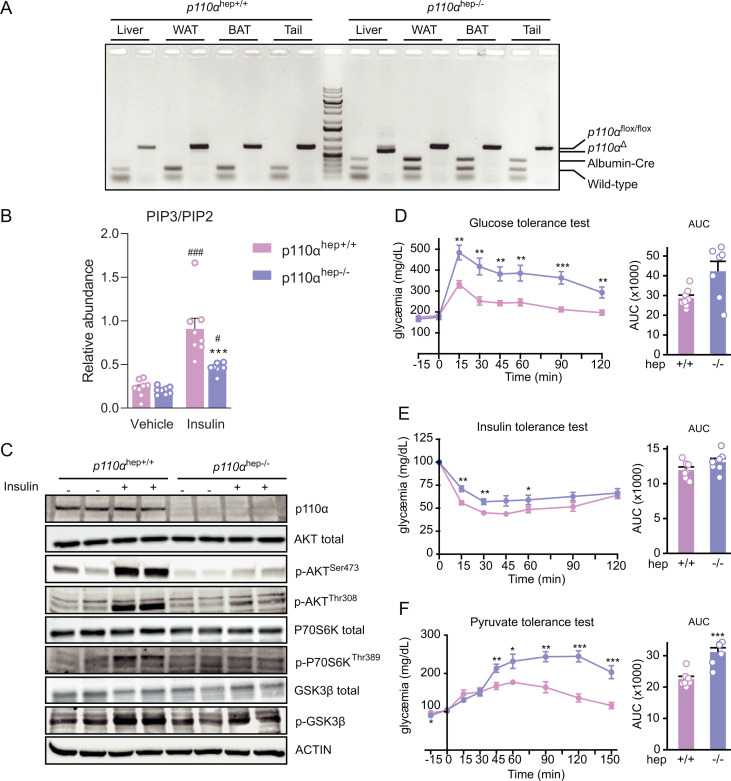
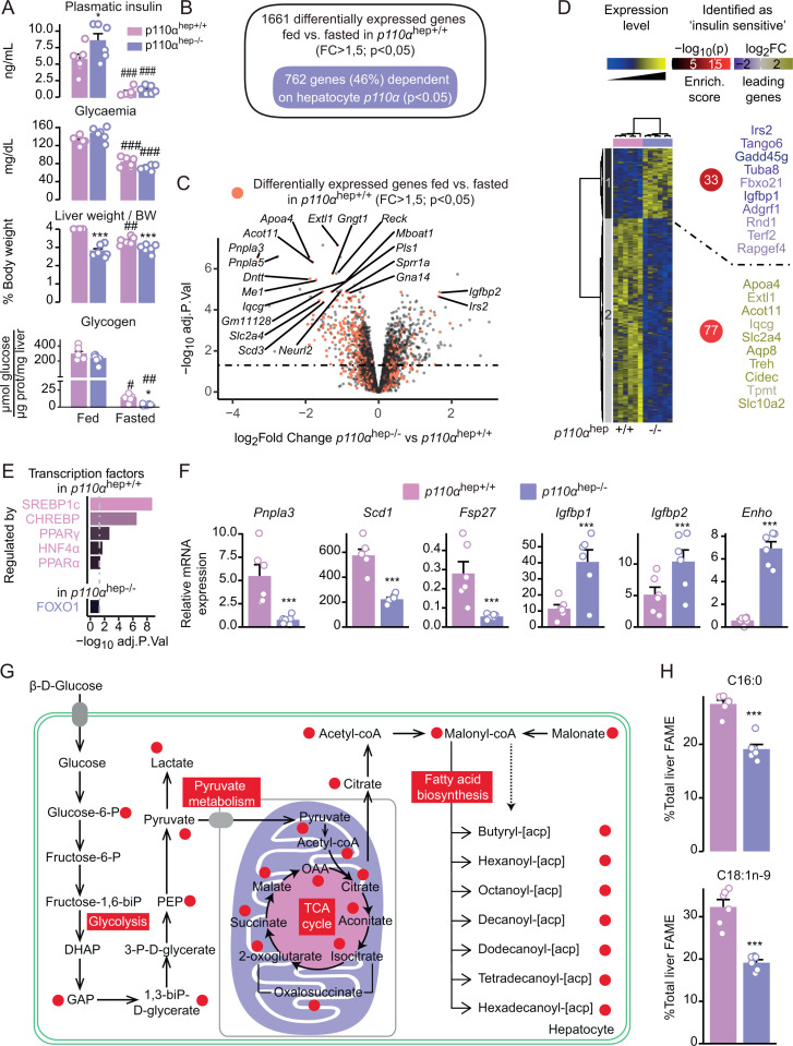
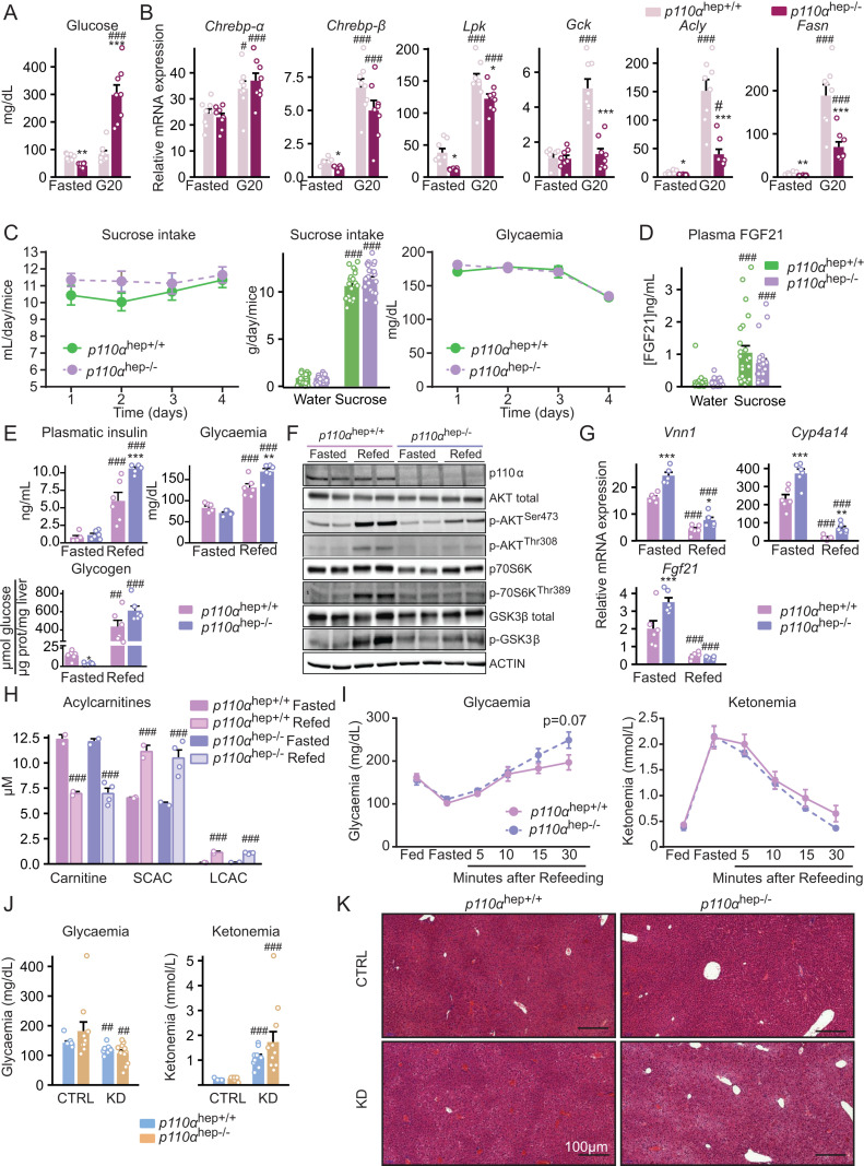
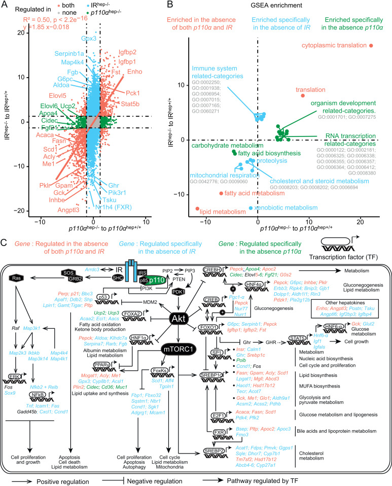
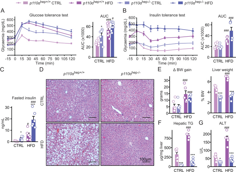
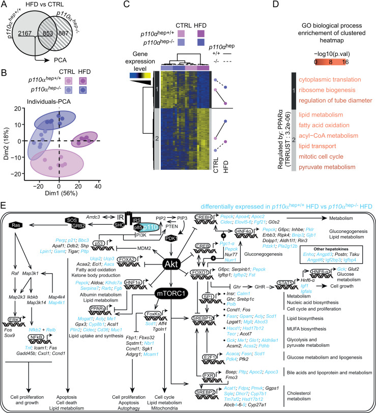
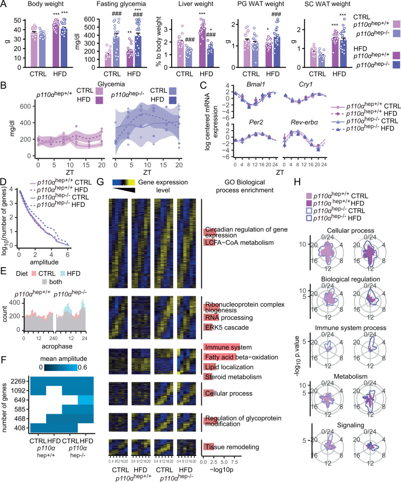
Insulin and other growth factors are key regulators of liver gene expression, including in metabolic diseases. Most of the phosphoinositide 3-kinase (PI3K) activity induced by insulin is considered to be dependent on PI3Kα. We used mice lacking p110α, the catalytic subunit of PI3Kα, to investigate its role in the regulation of liver gene expression in health and in metabolic dysfunction-associated steatotic liver disease (MASLD). The absence of hepatocyte PI3Kα reduced maximal insulin-induced PI3K activity and signaling, promoted glucose intolerance in lean mice and significantly regulated liver gene expression, including insulin-sensitive genes, in ad libitum feeding. Some of the defective regulation of gene expression in response to hepatocyte-restricted insulin receptor deletion was related to PI3Kα signaling. In addition, though PI3Kα deletion in hepatocytes promoted insulin resistance, it was protective against steatotic liver disease in diet-induced obesity. In the absence of hepatocyte PI3Kα, the effect of diet-induced obesity on liver gene expression was significantly altered, with changes in rhythmic gene expression in liver. Altogether, this study highlights the specific role of p110α in the control of liver gene expression in physiology and in the metabolic rewiring that occurs during MASLD.


Conflict of interest statement

The authors have declared that no competing interests exist.

Figures

Fig 1
Fig 1. Characterization of the hepatocyte-specific p110α knockout mouse model.
(A) PCR analysis of p110α floxed (p110αflox/flox), p110α deleted (p110αΔ), and Albumin-Cre (Albumin-Cre+/−) genes in control (p110αhep+/+) or liver knockout (p110αhep−/−) mice using genomic DNA from the liver, white adipose tissue (WAT), brown adipose tissue (BAT), and tail. (B) Liver PIP3/PIP2 ratio determined by LC/MS for p110αhep+/+ and p110αhep−/− mice that were fasted and then treated or not with insulin (5 U/kg) by vena cava injection (n = 8 mice/group). (C) Western blots of liver protein extracts evaluating the phosphorylation of AKT, p70S6K, and GSK3β in the mice in (B). (D–F) Glucose (D), insulin (E), and pyruvate (F) tolerance tests and corresponding AUC (n = 6 mice/genotype). Data information: In (B, D–F), data are presented as mean ± SEM. ##P ≤ 0.01 and ###P ≤ 0.005 for treatment effect; **P ≤ 0.01 and ***P ≤ 0.005 for genotype effect. The numerical values underlying the panels for this figure can be found in S1 Data.
Fig 2
Fig 2. Absence of p110α
-dependent signaling in hepatocytes affects liver growth, glucose homeostasis, and the regulation of liver gene expression. (A) Plasma insulin and glucose levels, relative liver weights and glycogen content in p110αhep+/+ and p110αhep−/− mice that were fed or fasted for 24 h and processed at ZT16 (n = 6 mice/group). (B) Venn diagram representing the genes regulated in a p110α-dependent manner in response to the nutritional status. (C) Volcano plot representing the differentially expressed genes in the liver of p110αhep−/− versus p110αhep+/+ fed mice. Red dots represent hepatic genes that are differentially expressed between fasting and feeding in p110hep+/+ mice. (D) Left: Heatmap illustrating microarray data for liver samples from fed p110αhep+/+ and p110αhep−/− mice. Right: Enrichment of insulin-sensitive genes as determined by [22]. The color of the circle and the gene names are relative to the percentage of all identified insulin-sensitive genes within a cluster and the P-value of the hypergeometric test. (E) Top list of the transcription factors up-regulated (top) and down-regulated (bottom) by p110α hepatocyte deletion as determined by TRRUST. (F) Relative expression of Pnpla3, Scd1, Fsp27, Igfbp1, Igfbp2, and Enho measured by qPCR in the livers of p110αhep+/+ and p110αhep−/− mice under fed conditions (n = 6 mice/group). (G) Profile of p110α-related metabolites based on iHepatocyte genome scale modeling visualized on an integrative representation of glycolysis, pyruvate metabolism, and fatty acid biosynthesis. Red circles represent metabolites significantly altered (P < 0.01) in the absence of p110α in the fed state. (H) Relative hepatic abundance of fatty acid methyl ester (FAME) C16:0 and C18:1n-9 determined by GC/MS. Data information: Data are presented as mean ± SEM. ***P ≤ 0.005 for genotype effect. The numerical values underlying the panels for this figure can be found in S2 Data which includes the full gene expression analysis for this figure in the sheet “Microarray Fig 2D”.
Fig 3
Fig 3. Hepatocyte p110α is required for insulin signaling but not for glucose and fatty acid sensing.
(A) p110αhep+/+ and p110αhep−/− mice were fasted or fed a chow diet supplemented with glucose (20%) in drinking water (n = 8 mice/group) and the plasma glucose levels measured. (B) Relative liver gene expression of Chrebp-α, Chrebp-β, Lpk, Gck, Acly, and Fasn measured by qPCR in the mice in (A). (C) Sucrose intake and plasma glucose levels measured daily during the sucrose preference test (10% sucrose in drinking water versus water, 4 days) (n = 23–24 mice/group). (D) Plasma FGF21 measured after 4 days of the sucrose preference test. (E) Plasma insulin and glucose levels, and liver glycogen content in p110αhep+/+ and p110αhep−/− mice fasted for 24 h and refed or not for 4 h with 20% glucose in drinking water (n = 6 mice/group). (F) Phosphorylation of AKT, p70S6K, and GSK3β determined by western blot in the mice in (E). (G) Relative liver expression of Vnn1, Cyp4a14, and Fgf21 mRNA measured by qPCR in the mice in (E). (H) Acylcarnitine levels measured in the blood of p110αhep+/+ and p110αhep−/− mice fasted for 24 h and refed or not for 4 h with 20% glucose in drinking water (n = 3 mice/group). (I) Blood glucose and ketone levels measured in p110αhep+/+ and p110αhep−/− mice successively fed, fasted (24 h), and refed for 5, 10, 15, or 30 min (n = 6 mice/group). (J) Blood glucose and ketone levels measured in plasma from p110αhep+/+ and p110αhep−/− mice fed a CTRL diet or ketogenic diet (KD) (n = 9–11 mice/group). (K) Representative pictures of H/E staining of liver sections from the mice in (J). Scale bar, 100 µM. Data information: In (A–D, H, J), data are presented as mean ± SEM. #P ≤ 0.05, ##P ≤ 0.01, and ###P ≤ 0.005 for glucose (A, B) or sucrose effect (C, D), or diet (J) or nutritional status effect (H); *P ≤ 0.05, **P ≤ 0.01, and ***P ≤ 0.005 for genotype effect. The numerical values underlying the panels for this figure can be found in S3 Data.
Fig 4
Fig 4. Hepatocyte p110α-mediated regulation of gene expression related to IR-dependent and IR-independent pathways.
(A) Correlation plot of data extracted from two distinct microarray experiments. The values used for correlation are –log10 adjusted P-value of the comparison indicated on the axis, weighted by the sign of the corresponding log2FC of expression for each gene. (B) Correlation plot of gene set enrichment over genes presented in (A). The values used to plot the significant categories are the –log10 adjusted P-value of the comparison indicated on the axis, weighted by the sign of the corresponding normalized enrichment score. A manual annotation summarizes related GO categories when they cluster in the same area of the plot. (C) Schematic overview of insulin-responsive transcription factors and downstream-regulated genes dependent on IR, p110α, or both. Genes in blue are regulated in absence of IR but not in absence of p110α. Genes in green are regulated in absence of p110α but not IR. Genes in orange are regulated by both IR and p110α.
Fig 5
Fig 5. Hepatocyte p110α deficiency disconnects hepatic steatosis from diabetes.
(A, B) Glucose (A) and insulin (B) tolerance tests and corresponding AUC for 12-week-old p110αhep+/+ and p110αhep−/− mice fed a chow diet (CTRL) or HFD for 12 weeks (n = 6 mice/group). (C) Plasma insulin levels in 24 h-fasted p110αhep+/+ and p110αhep−/− mice fed CTRL or HFD for 12 weeks. (D) Representative pictures of H/E staining of liver sections. Scale bar, 100 µm. (E) Body weight gain and liver weight at the end of the experiment in mice from (A). (F) Liver content in triglycerides. (G) Plasma ALT levels. Data information: In all graphs, data are presented as mean ± SEM. #P ≤ 0.05 and ###P ≤ 0.005 for diet effect; *P ≤ 0.05 and ***P ≤ 0.005 for genotype effect. The numerical values underlying the panels for this figure can be found in S4 Data.
Fig 6
Fig 6. Hepatocyte signaling dependent on p110α regulates HFD-mediated changes in liver gene expression profile.
(A) Euler diagram representing the number of HFD-sensitive genes determined by microarray for each genotype of 12-week-old p110αhep+/+ and p110αhep−/− mice fed a chow diet (CTRL) or HFD for 12 weeks (n = 6 mice/group). (B) Principal component analysis (PCA) of genes selected in (A) that were regulated by HFD in both genotypes. (C) Heatmap with hierarchical clustering of genes presenting a >75% correlation to dimension 1 of the PCA in (B) and mean z-score for each group within each cluster. (D) Enrichment in GO Biological Process categories on clusters determined in (C) and TRRUST enrichment. (E) Schematic overview of insulin-responsive transcription factors and downstream-regulated genes dependent on or independent of p110α during HFD-induced obesity. Genes in blue are differentially expressed in p110αhep−/− mice during obesity. Genes in black are similarly expressed in p110αhep+/+ and p110αhep−/− mice fed an HFD. The full gene expression analysis for this figure can be found in S4 Data, in the sheet “Microarray Fig 6A–C”.
Fig 7
Fig 7. p110α dependent signaling is important for the rewiring of gene expression that occurs during obesity.
(A) Body weight, fasting plasma glucose levels, and liver, perigonadal, and subcutaneous adipose tissue relative weights in 12-week-old p110αhep+/+ and p110αhep−/− mice fed a chow diet (CTRL) or HFD diet for 12 weeks (n = 12–18/group). Data are presented as mean ± SEM. #P ≤ 0.05 and ###P ≤ 0.005 for diet effect; *P ≤ 0.05 and ***P ≤ 0.005 for genotype effect. (B) Plasma glucose levels in p110αhep+/+ (left) and p110αhep−/− (right) mice fed a CTRL or HFD diet around the clock (ZT0 to ZT20). (C) Relative hepatic mRNA expression levels of core clock genes and clock-controlled genes from qPCR analyzed by the Drylm function (DryR package). (D, E) Analysis of circadian gene expression from microarray analysis of the liver, including the cumulative number of rhythmic genes (D) and phase distribution of rhythmic genes (E). (F) Representation of the six rhythmic models identified by dryR that present a significant result in hypergeometric testing for at least one of the first level GO Biological Process categories. Each line represents a model, and each column an experimental group. When a group has no rhythmic parameter, the corresponding tile remains empty. When two groups share the same rhythmic parameters, their tiles are colored proportionally to the mean amplitude. (G) Heatmap of the models selected in (F). The heatmap rows, one per gene, are sorted by acrophase. Data are scaled by row. The heatmap columns are sorted by hour (ZT0, 4, 8, 12, 16, 20). The right panel represents the GO biological process enrichment for each transcriptomic rhythmic profile. (H) Functional enrichment around the clock. Enrichment scores for the indicated functional terms are represented by the radial coordinate at the indicated time point. p110αhep+/+ mice are represented on the left and p110αhep−/− mice on the right. The numerical values underlying the panels for this figure can be found in S5 Data. The full gene expression analysis for this figure can be found in S5 Data, in the sheet “DryR parameters”.

Similar articles

References

    1. Hawkins PT, Anderson KE, Davidson K, Stephens LR. Signalling through Class I PI3Ks in mammalian cells. Biochem Soc Trans. 2006;34(Pt 5):647–62. doi: 10.1042/BST0340647 . - DOI - PubMed
    1. White MF, Kahn CR. Insulin action at a molecular level—100 years of progress. Mol Metab. 2021;52:101304. doi: 10.1016/j.molmet.2021.101304 . - DOI - PMC - PubMed
    1. Roden M, Shulman GI. The integrative biology of type 2 diabetes. Nature. 2019;576(7785):51–60. doi: 10.1038/s41586-019-1797-8 . - DOI - PubMed
    1. Titchenell PM, Lazar MA, Birnbaum MJ. Unraveling the regulation of hepatic metabolism by insulin. Trends Endocrinol Metab. 2017;28(7):497–505. doi: 10.1016/j.tem.2017.03.003 . - DOI - PMC - PubMed
    1. Batista TM, Cederquist CT, Kahn CR. The insulin receptor goes nuclear. Cell Res. 2019;29(7):509–11. doi: 10.1038/s41422-019-0185-0 . - DOI - PMC - PubMed

MeSH terms

LinkOut - more resources