Brown-Sequard syndrome caused by posterior full-endoscopic cervical discectomy: A case report
- PMID: 40228252
- PMCID: PMC11999450
- DOI: 10.1097/MD.0000000000042007
Brown-Sequard syndrome caused by posterior full-endoscopic cervical discectomy: A case report
Abstract
Background: Posterior full-endoscopic cervical discectomy (PFECD) is an effective and safe technique for cervical radiculopathy. The primary complications of PFECD include temporary nerve root paralysis and dural rupture, while spinal cord damage is exceedingly rare. This study describes a rare case of Brown-Sequard syndrome (BSS) occurring following PFECD and investigates its potential etiologies and pathomechanisms associated with this procedure.
Methods: Notes and images were reviewed and the relevant literature was analyzed.
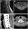
Results: A 50-year-old woman underwent PFECD for cervical radiculopathy. The patient reported substantial alleviation of radicular pain symptoms on the first postoperative day. On the third postoperative day, the patient exhibited acute-onset weakness in the left lower limb, along with diminished pinprick and temperature sensation in the right limb. Cervical spine magnetic resonance imaging demonstrated a newly developed T2 hyperintensity at the C5 spinal cord level. BSS was confirmed based on correlating imaging findings with clinical signs. Following the comprehensive treatment of rehabilitation and pharmacological therapy, the patient's neurological deficits symptoms gradually improvement. At the 6-month follow-up, the patient's symptoms resolved entirely, and the T2 hypersignal diminished markedly on repeat magnetic resonance imaging.
Conclusion: This study represents the first case of BSS following PFECD. We emphasize that although the PFECD technique is safe and effective, meticulous surgical technique-particularly in foraminal decompression-is critical to avoid iatrogenic spinal cord injury.
Keywords: Brown; Sequard syndrome; endoscopic cervical discectomy; posterior full; spinal cord injury.
Copyright © 2025 the Author(s). Published by Wolters Kluwer Health, Inc.
Conflict of interest statement
The author have no conflicts of interest to disclose.
Figures


Similar articles
-
Brown-Séquard syndrome after a gun shot wound to the cervical spine: a case report.Spine J. 2013 Dec;13(12):e1-5. doi: 10.1016/j.spinee.2013.06.093. Epub 2013 Sep 17. Spine J. 2013. PMID: 24051332
-
Brown-Sèquard syndrome produced by C3-C4 cervical disc herniation: a case report and review of the literature.Spine (Phila Pa 1976). 2008 Apr 20;33(9):E279-82. doi: 10.1097/BRS.0b013e31816c835d. Spine (Phila Pa 1976). 2008. PMID: 18427307 Review.
-
Brown-Sèquard syndrome produced by cervical disc herniation: report of two cases and review of the literature.Spine J. 2003 Nov-Dec;3(6):530-3. doi: 10.1016/s1529-9430(03)00078-0. Spine J. 2003. PMID: 14609700 Review.
-
Cervical stenosis presenting with acute Brown-Séquard syndrome: case report.Spine (Phila Pa 1976). 2005 Aug 15;30(16):E481-3. doi: 10.1097/01.brs.0000174284.08440.1e. Spine (Phila Pa 1976). 2005. PMID: 16103843
-
Brown Sequard syndrome in a patient with Klippel-Feil syndrome following minor trauma: a case report and literature review.BMC Musculoskelet Disord. 2023 Sep 11;24(1):722. doi: 10.1186/s12891-023-06760-9. BMC Musculoskelet Disord. 2023. PMID: 37697343 Free PMC article. Review.
References
-
- Deng ZL, Chu L, Chen L, Yang JS. Anterior transcorporeal approach of percutaneous endoscopic cervical discectomy for disc herniation at the C4–C5 levels: a technical note. Spine J. 2016;16:659–66. - PubMed
-
- Wu PF, Li YW, Wang B, Jiang B, Tu ZM, Lv GH. Posterior cervical foraminotomy via full-endoscopic versus microendoscopic approach for radiculopathy: a systematic review and meta-analysis. Pain Physician. 2019;22:41–52. - PubMed
-
- Kobayashi N, Asamoto S, Doi H, Sugiyama H. Brown-Sèquard syndrome produced by cervical disc herniation: report of two cases and review of the literature. Spine J. 2003;3:530–3. - PubMed
Publication types
MeSH terms
Grants and funding
LinkOut - more resources
Full Text Sources
Medical
Miscellaneous

