Traumatic basal ganglia hemorrhage: illustrative cases
- PMID: 40228406
- PMCID: PMC12001058
- DOI: 10.3171/CASE24625
Traumatic basal ganglia hemorrhage: illustrative cases
Abstract
Background: Traumatic basal ganglia hemorrhage (TBGH) is rare. The most common mechanism of injury is road traffic accidents. In this case series, the authors discuss the clinical course of 5 patients with TBGH with different outcomes.
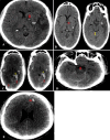
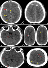
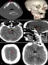
Observations: The internal capsule is the most commonly involved site, which was noted in 3 of the 5 cases reported here. The size of the TBGHs ranged from 1.02 cm to 2.61 cm. All patients had at least 1 additional site of bleeding. One patient had zygomatic, maxillary, and mandibular fractures, while another patient had a mandibular fracture. Two of the 5 patients died. These 2 patients had Glasgow Coma Scale (GCS) scores of 3 and 4, and their pupils were not reactive to light after resuscitation and loading with mannitol.
Lessons: CT findings in TBGHs differ from those in spontaneous hemorrhages in that gray-white matter junction contusions and ventral or dorsolateral brainstem contusions are more commonly observed in the former. Compared with other fractures, mandibular fractures are more commonly associated with TBGH. Conservative treatment is a valid approach for managing patients with TBGH. The overall prognosis of patients with TBGH is poor, and the highest mortality rates are seen in patients with low GCS scores and absent pupillary light reactions. https://thejns.org/doi/10.3171/CASE24625.
Keywords: case series; diffuse axonal injury; head injury; road traffic accident; traumatic basal ganglia hemorrhage.
Figures


Similar articles
-
Bilateral basal ganglia bleed in traumatic brain injury: A case report.J Pak Med Assoc. 2020 Oct;70(10):1854-1856. doi: 10.5455/JPMA.36070. J Pak Med Assoc. 2020. PMID: 33159769
-
Clinical outcomes and prognostic factors of traumatic basal ganglia hematomas: A 4-year single-center study.Surg Neurol Int. 2023 Jul 21;14:251. doi: 10.25259/SNI_411_2023. eCollection 2023. Surg Neurol Int. 2023. PMID: 37560578 Free PMC article.
-
Prognostic Significance of Magnetic Resonance Imaging in Detecting Diffuse Axonal Injuries: Analysis of Outcomes and Review of Literature.Neurol India. 2022 Nov-Dec;70(6):2371-2377. doi: 10.4103/0028-3886.364066. Neurol India. 2022. PMID: 36537418 Review.
-
Large traumatic basal ganglia hematoma: surgical treatment versus conservative management.J Neurosurg Sci. 2020 Apr;64(2):154-157. doi: 10.23736/S0390-5616.16.03830-3. Epub 2016 Nov 17. J Neurosurg Sci. 2020. PMID: 27854112
-
Intracerebral hemorrhage due to cerebral amyloid angiopathy after head injury: Report of a case and review of the literature.Neuropathology. 2016 Dec;36(6):566-572. doi: 10.1111/neup.12308. Epub 2016 May 5. Neuropathology. 2016. PMID: 27145894 Review.
References
-
- Nabika S Kiya K Satoh H Mizoue T Oshita J Kondo H.. Ischemia of the internal capsule due to mild head injury in a child. Pediatr Neurosurg. 2007;43(4):312-315. - PubMed
-
- Boto GR Lobato RD Rivas JJ Gomez PA de la Lama A Lagares A.. Basal ganglia hematomas in severely head injured patients: clinicoradiological analysis of 37 cases. J Neurosurg. 2001;94(2):224-232. - PubMed
-
- Haas DC Pineda GS Lourie H.. Juvenile head trauma syndromes and their relationship to migraine. Arch Neurol. 1975;32(11):727-730. - PubMed
LinkOut - more resources
Full Text Sources

