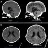
Reversible abducens nerve palsy following cranial vault expansion in the setting of multisutural craniosynostosis: illustrative case
- PMID: 40228410
- PMCID: PMC12001060
- DOI: 10.3171/CASE24762
Reversible abducens nerve palsy following cranial vault expansion in the setting of multisutural craniosynostosis: illustrative case
Abstract
Background: Posterior cranial vault distraction osteogenesis (PVDO) is a commonly used cranial expansion procedure in infants and children with syndromic craniosynostosis. To date, there have been no reports of cranial nerve (CN) palsies in patients undergoing univector PVDO.
Observations: In this article, the authors describe the case of a 27-month-old female with Muenke syndrome who underwent long-distance (> 30 mm) PVDO and developed bilateral abducens nerve (CN VI) palsy after 40 mm of distraction. Following partial reversal of the distraction during the activation phase, the authors observed complete resolution of this palsy.
Lessons: This report demonstrates that CN palsies are a potential complication for which the patient should be monitored, even when undergoing univector PVDO. Most notably, this report illustrates that a gradual reduction in the distraction distance can result in complete resolution of a CN VI palsy while also maintaining a significant degree of intracranial expansion. https://thejns.org/doi/10.3171/CASE24762.
Keywords: CN VI; abducens palsy; cranial nerve VI; craniosynostosis; lateral rectus palsy; posterior cranial vault distraction osteogenesis.
Figures


Similar articles
-
Posterior vault distraction osteogenesis: indications and expectations.Childs Nerv Syst. 2021 Oct;37(10):3119-3125. doi: 10.1007/s00381-021-05118-7. Epub 2021 Mar 20. Childs Nerv Syst. 2021. PMID: 33743044
-
Complete posterior cranial vault distraction osteogenesis to correct Chiari malformation type I associated with craniosynostosis.J Neurosurg Pediatr. 2021 Dec 17;29(3):298-304. doi: 10.3171/2021.10.PEDS21443. Print 2022 Mar 1. J Neurosurg Pediatr. 2021. PMID: 34920435
-
Onset and Resolution of Chiari Malformations and Hydrocephalus in Syndromic Craniosynostosis following Posterior Vault Distraction.Plast Reconstr Surg. 2019 Oct;144(4):932-940. doi: 10.1097/PRS.0000000000006041. Plast Reconstr Surg. 2019. PMID: 31568307
-
Craniosynostosis: Posterior Cranial Vault Remodeling.Clin Plast Surg. 2021 Jul;48(3):455-471. doi: 10.1016/j.cps.2021.03.001. Epub 2021 May 11. Clin Plast Surg. 2021. PMID: 34051898 Review.
-
Posterior cranial vault distraction osteogenesis: A systematic review.J Oral Biol Craniofac Res. 2022 Nov-Dec;12(6):823-832. doi: 10.1016/j.jobcr.2022.09.009. Epub 2022 Sep 16. J Oral Biol Craniofac Res. 2022. PMID: 36186267 Free PMC article. Review.
References
-
- Derderian CA Wink JD McGrath JL Collinsworth A Bartlett SP Taylor JA.. Volumetric changes in cranial vault expansion: comparison of fronto-orbital advancement and posterior cranial vault distraction osteogenesis. Plast Reconstr Surg. 2015;135(6):1665-1672. - PubMed
-
- Li J Gerety PA Xu W Bartlett SP Taylor JA.. A perioperative risk comparison of posterior vault distraction osteogenesis in an older pediatric population. J Craniofac Surg. 2016;27(5):1165-1169. - PubMed
-
- Taylor JA Derderian CA Bartlett SP Fiadjoe JE Sussman EM Stricker PA.. Perioperative morbidity in posterior cranial vault expansion: distraction osteogenesis versus conventional osteotomy. Plast Reconstr Surg. 2012;129(4):674e-680e. - PubMed
-
- Greives MR Ware BW Tian AG Taylor JA Pollack IF Losee JE.. Complications in posterior cranial vault distraction. Ann Plast Surg. 2016;76(2):211-215. - PubMed
-
- Humphries LS, Zapatero ZD, Vu GH.Ten years of posterior cranial vault expansion by means of distraction osteogenesis: an update and critical evaluation. Plast Reconstr Surg. 2022;150(2):379-391. - PubMed
LinkOut - more resources
Full Text Sources

