

Intracerebellar extension of occipital scalp dermoid after initial resection: illustrative case
- PMID: 40228411
- PMCID: PMC12001065
- DOI: 10.3171/CASE2548
Intracerebellar extension of occipital scalp dermoid after initial resection: illustrative case
Abstract
Background: Dermoid cysts are benign ectodermal growths that commonly form at cranial sutures. Although benign, resection is often recommended to prevent intracranial extension as the cyst grows. The prognosis after resection is very good, provided a complete resection is possible.
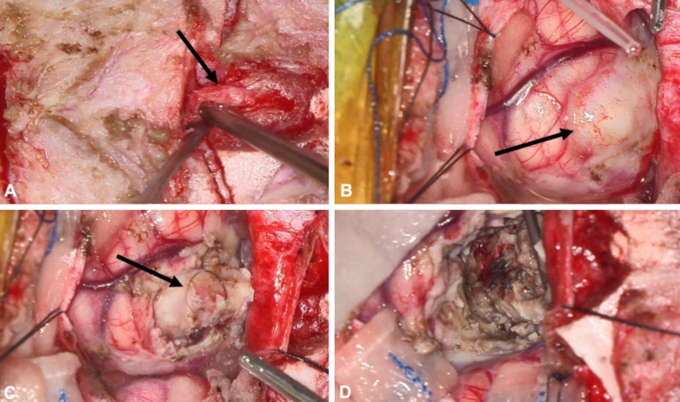
Observations: The authors present the case of a 10-month-old male patient with an occipital scalp dermoid cyst with intracranial extension who underwent subtotal resection and later presented with cerebellar extension.
Lessons: Scalp dermoid cysts with intradiploic extension have a significant risk of intracranial extension. Thus, care must be taken to achieve total resection in the first attempt or provide close follow-up before intracranial extension develops. https://thejns.org/doi/10.3171/CASE2548.
Keywords: hydrocephalus; intracranial dermoid cyst; pediatrics.
Figures


Similar articles
-
A Rare Presentation of Occipital Dermoid Cyst with Intracranial Extension and Secondary Infection: Case Report and Follow-Up.J Neurol Surg Rep. 2024 Apr 8;85(2):e39-e42. doi: 10.1055/a-2287-2108. eCollection 2024 Apr. J Neurol Surg Rep. 2024. PMID: 38596231 Free PMC article.
-
Dermoid and Epidermoid Cysts of Scalp: Case Series of 234 Consecutive Patients.World Neurosurg. 2018 Dec;120:119-124. doi: 10.1016/j.wneu.2018.08.197. Epub 2018 Sep 3. World Neurosurg. 2018. PMID: 30189303 Review.
-
Intradiploic dermoid cyst of the lateral frontotemporal skull: case report and review of the literature.Pediatr Neurosurg. 2013;49(4):232-5. doi: 10.1159/000363329. Epub 2014 Jul 9. Pediatr Neurosurg. 2013. PMID: 25012262 Review.
-
Dermoid Cyst of the Prepontine Cistern and Meckel's Cave: Illustrative Case and Systematic Review.J Neurol Surg B Skull Base. 2018 Apr;79(2):139-150. doi: 10.1055/s-0037-1604332. Epub 2017 Aug 11. J Neurol Surg B Skull Base. 2018. PMID: 29868318 Free PMC article.
-
Malignant transformation of a cerebral dermoid cyst into a squamous cell carcinoma with malignant intraperitoneal spreading along a ventriculoperitoneal shunt: illustrative case.J Neurosurg Case Lessons. 2022 Oct 17;4(16):CASE2254. doi: 10.3171/CASE2254. Print 2022 Oct 17. J Neurosurg Case Lessons. 2022. PMID: 36254352 Free PMC article.
References
-
- Prior A, Anania P, Pacetti M.Dermoid and epidermoid cysts of scalp: case series of 234 consecutive patients. World Neurosurg. 2018;120:119-124. - PubMed
-
- Nakajima K Korekawa A Nakano H Sawamura D.. Subcutaneous dermoid cysts on the eyebrow and neck. Pediatr Dermatol. 2019;36(6):999-1001. - PubMed
-
- Julapalli MR Cohen BA Hollier LH Metry DW.. Congenital, ill-defined, yellowish plaque: the nasal dermoid. Pediatr Dermatol. 2006;23(6):556-559. - PubMed
-
- Orozco-Covarrubias L Lara-Carpio R Saez-De-Ocariz M Duran-McKinster C Palacios-Lopez C Ruiz-Maldonado R.. Dermoid cysts: a report of 75 pediatric patients. Pediatr Dermatol. 2013;30(6):706-711. - PubMed
LinkOut - more resources
Full Text Sources
