

Intracranial malignant melanotic nerve sheath tumor: illustrative case
- PMID: 40228412
- PMCID: PMC12001059
- DOI: 10.3171/CASE24473
Intracranial malignant melanotic nerve sheath tumor: illustrative case
Abstract
Background: Melanotic schwannoma accounts for 1% of all nerve sheath tumors. These tumors were considered benign, but recent studies have shown their malignant propensity. They pose a diagnostic challenge given the rarity of the tumor.
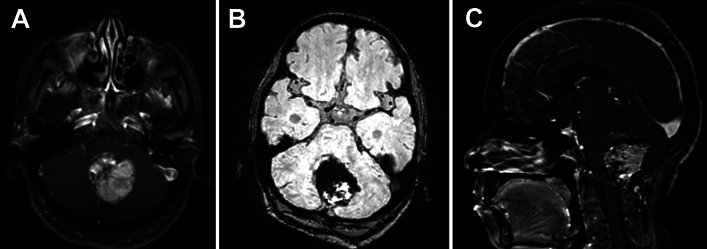
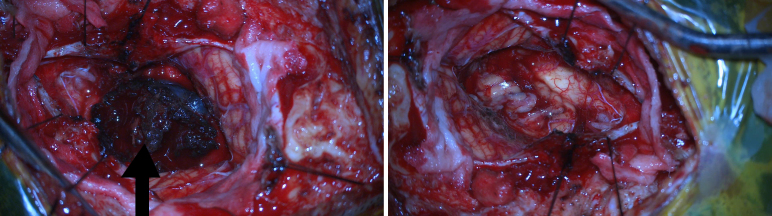
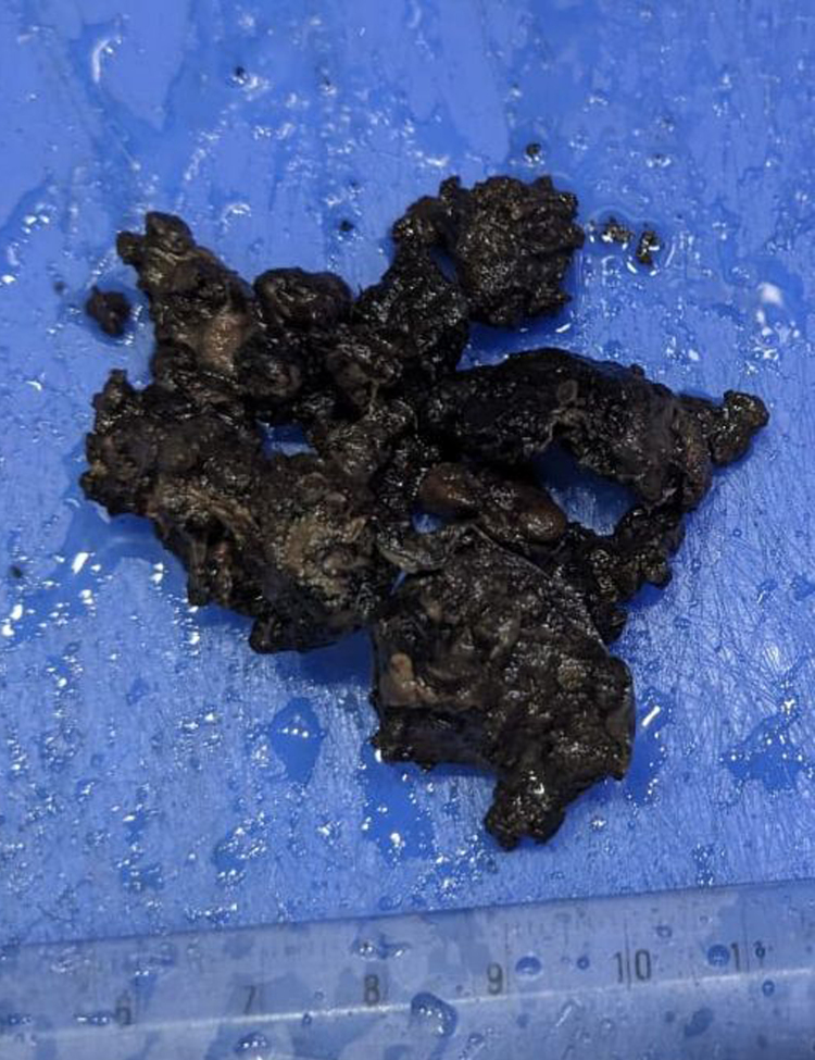
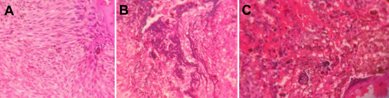
Observations: The authors report a case of a 42-year-old woman who presented with headaches and a history of frequent falls for the past year. Brain MRI revealed a lesion in the posterior fossa, effacing the median aperture and cisterna magna, which was hyperintense on T1-weighted imaging and isointense on T2-weighted imaging, with significant blooming on susceptibility-weighted imaging. The patient underwent a midline suboccipital craniotomy and gross-total resection of the tumor. Histopathological examination revealed a malignant melanotic nerve sheath tumor (MMNST) with psammoma bodies and necrosis, indicating a poor prognosis.
Lessons: Melanotic schwannomas have a malignant propensity despite their benign morphology. Immunohistochemical analysis helps confirm the diagnosis of melanotic schwannoma. This is the 21st documented case of an intracranial MMNST, and, given the rarity of the tumor, there is scope for further research and studies on the role of radiotherapy in the management of these tumors. https://thejns.org/doi/10.3171/CASE24473.
Keywords: case report; intracranial; melanotic schwannoma; necrosis.
Figures
Similar articles
-
Extradural malignant melanotic nerve sheath tumor of the lumbosacral spine: a diagnostic and surgical challenge.Ann Med Surg (Lond). 2025 May 12;87(6):3940-3944. doi: 10.1097/MS9.0000000000003327. eCollection 2025 Jun. Ann Med Surg (Lond). 2025. PMID: 40486650 Free PMC article.
-
Malignant melanotic nerve sheath tumors in the spinal canal of psammomatous and non-psammomatous type: Two case reports.World J Clin Cases. 2022 Aug 26;10(24):8735-8741. doi: 10.12998/wjcc.v10.i24.8735. World J Clin Cases. 2022. PMID: 36157803 Free PMC article.
-
Sporadic spinal psammomatous malignant melanotic nerve sheath tumor: A case report and literature review.Front Oncol. 2023 Feb 24;13:1100532. doi: 10.3389/fonc.2023.1100532. eCollection 2023. Front Oncol. 2023. PMID: 36910634 Free PMC article.
-
A para-aortic malignant melanotic nerve sheath tumor mimicking a gastrointestinal stromal tumor: a rare case report and review of literature.BMC Surg. 2022 Jul 28;22(1):293. doi: 10.1186/s12893-022-01727-4. BMC Surg. 2022. PMID: 35902891 Free PMC article. Review.
-
Dumbbell-shaped C-2 psammomatous melanotic malignant schwannoma. Case report and review of the literature.J Neurosurg Spine. 2007 Jun;6(6):591-9. doi: 10.3171/spi.2007.6.6.14. J Neurosurg Spine. 2007. PMID: 17561752 Review.
References
-
- Mahato D Vivas-Buitrago T Gassie K Jentoft M Tavanaiepour D Quiñones-Hinojosa A.. Intracranial melanotic schwannomas: a rare variant with unusual adherent features. J Neurooncol. 2018;136(2):299-306. - PubMed
-
- Torres-Mora J Dry S Li X Binder S Amin M Folpe AL.. Malignant melanotic Schwannian tumor: a clinicopathologic, immunohistochemical, and gene expression profiling study of 40 cases, with a proposal for the reclassification of “melanotic schwannoma”. Am J Surg Pathol. 2014;38(1):94-105. - PubMed
-
- Malhotra A Rao S Santhoshkumar R Muralidharan N Mitra S Shetty S.. Pigmented ependymoma of the fourth ventricle—a curious entity: report of a rare case with review of literature. Int J Surg Pathol. 2021;29(1):80-84. - PubMed
LinkOut - more resources
Full Text Sources
Miscellaneous
