. 2025 Jun;40(3):340-348.
doi: 10.1016/j.virs.2025.04.005. Epub 2025 Apr 12.

Viral pseudo-enzyme facilitates KSHV lytic replication via suppressing PFAS-mediated RTA deamidation

Affiliations

Viral pseudo-enzyme facilitates KSHV lytic replication via suppressing PFAS-mediated RTA deamidation

Yang Xu et al. Virol Sin. 2025 Jun.

Abstract

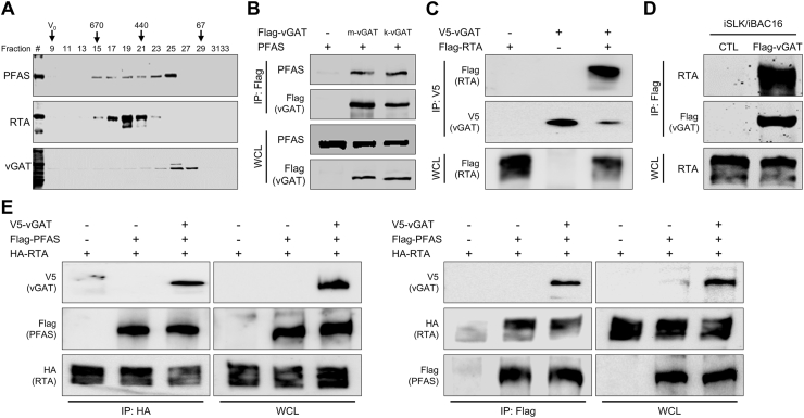
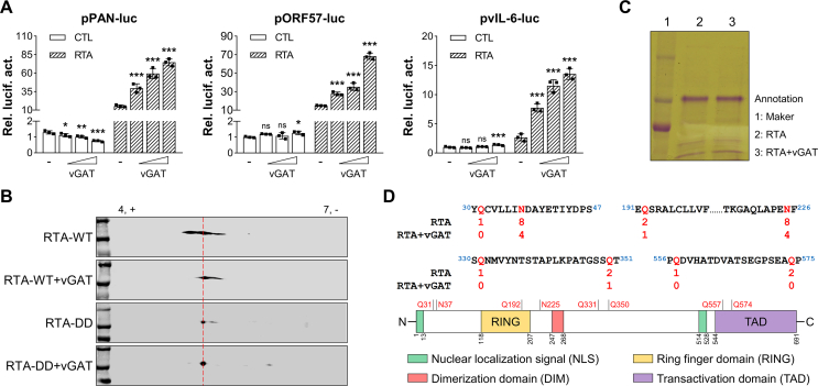
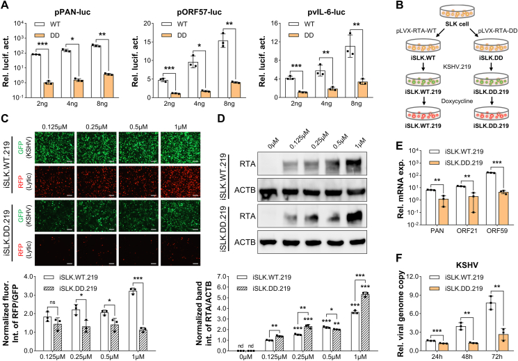
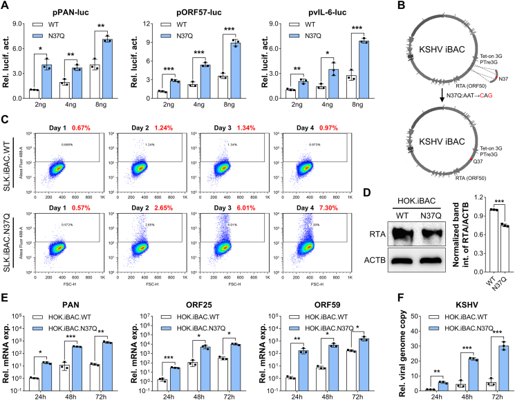
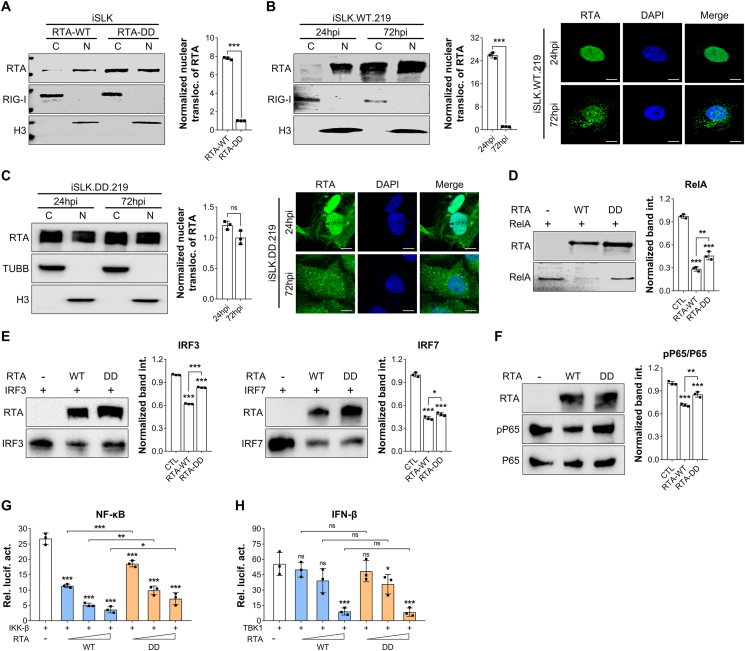
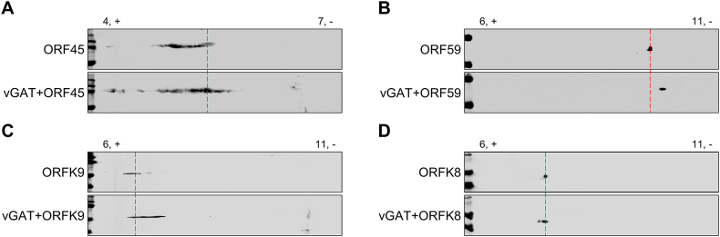
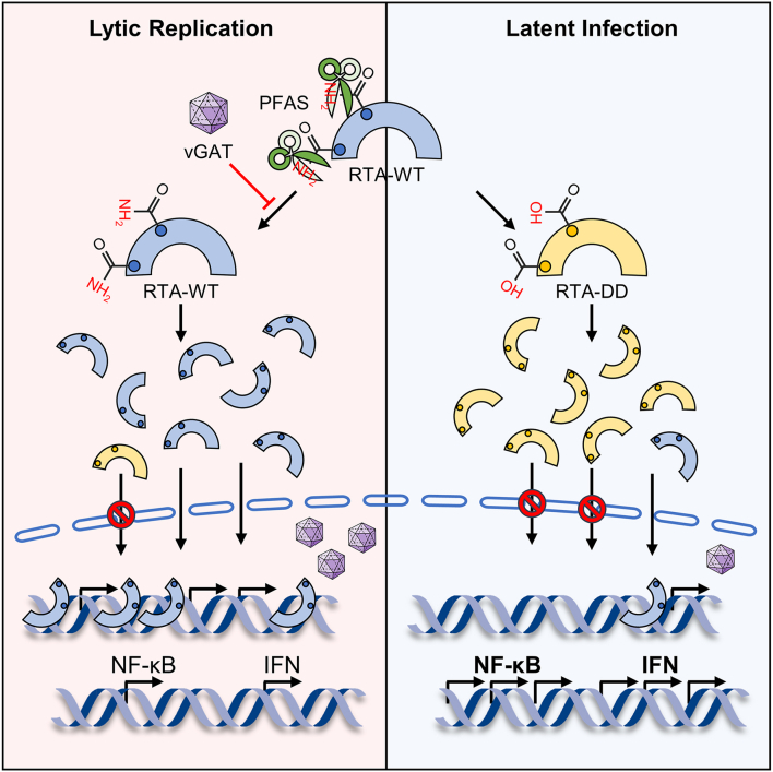
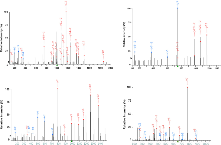
Deamidation, a type of post-translational modification commonly considered a hallmark of protein "aging" and function decay, is increasingly recognized for its pivotal role in regulating biological processes and viral infection. Our previous study has demonstrated that the deamidation of replication and transcription activator (RTA), a master regulator of ubiquitous and oncogenic Kaposi's sarcoma-associated herpesvirus (KSHV), mediated by phosphoribosylformylglycinamidine synthetase (PFAS), hinders its nuclear import and transcriptional activity. Here we report that the viral glutamine amidotransferase (vGAT) pseudo-enzyme is exploited to facilitate KSHV lytic infection by inhibiting RTA deamidation. To be more specific, vGAT interacts with both RTA and cellular PFAS, and inhibits PFAS-mediated RTA deamidation, thus facilitating RTA nuclear localization and suppressing nuclear factor-kappa B (NF-κB) signaling activation, as well as augmenting RTA-mediated transcriptional activation of viral open reading frames (ORFs). In addition, vGAT appears to regulate the deamidation process of several viral ORFs of KSHV. Collectively, these findings unveil that a viral pseudo-enzyme is exploited to enhance viral infection via deamidation regulation.

Keywords: Deamidation; Kaposi's sarcoma-associated herpesvirus (KSHV); transcription activator (RTA); viral glutamine amidotransferase (vGAT).


Conflict of interest statement

Conflict of interest The authors declare that they have no conflict of interest.

Figures

Fig. 1
Fig. 1
vGAT interacts with both RTA and cellular PFAS. A 293T cells were infected with KSHV (multiplicity of infection, MOI ​= ​5) for 24 ​h. Purified PFAS, RTA, and vGAT were analyzed by gel filtration. B 293T cells were transfected with plasmids expressing MHV68 (m-vGAT) or KSHV (k-vGAT) GAT and plasmids expressing PFAS. Whole-cell lysates (WCLs) were prepared and immunoprecipitation was performed. Precipitated proteins and WCLs were analyzed by immunoblotting with indicated antibodies. C 293T cells were transfected with indicated plasmids. WCLs were prepared and immunoprecipitation was performed. Precipitated proteins and WCLs were analyzed by immunoblotting with indicated antibodies. D iSLK/iBAC16 ​cells were induced with doxycycline (1 ​μg/mL) for 48 ​h. WCLs were prepared and immunoprecipitation was performed. Precipitated proteins and WCLs were analyzed by immunoblotting with indicated antibodies. E 293T cells were transfected with indicated plasmids. WCLs were prepared and immunoprecipitation was performed. Precipitated proteins and WCLs were analyzed by immunoblotting with indicated antibodies.
Fig. 2
Fig. 2
vGAT inhibits the deamidation of RTA. A 293T cells were transfected with indicated reporter cocktails and different dose of plasmids expressing vGAT (1 ​ng, 10 ​ng, and 50 ​ng) without or with RTA (1 ​ng). vGAT mediated RTA-dependent transcriptional activation of the indicated promoters was determined by luciferase reporter assay. Data presented as mean ​± ​standard deviations. Statistical analyses were performed using one-way analysis of variance (ANOVA). Statistical significance was set at ∗P ​< ​0.05, ∗∗P ​< ​0.01, ∗∗∗P ​< ​0.001, while “ns” indicated “not significant”. B 293T cells were transfected with plasmids expressing wild type RTA (RTA-WT) or RTA deamidation mutant (RTA-DD) without or with vGAT. WCLs were analyzed by two-dimensional gel electrophoresis and immunoblotting. C 293T cells were transfected with plasmids expressing RTA without or with vGAT. RTA was purified via single step affinity chromatography using anti-FLAG M2 agarose and eluted with FLAG peptide (0.2 ​mg/mL). Proteins remained on beads were analyzed by SDS-PAGE and Coomassie staining. The RTA band was excised and analyzed by tandem mass spectrometry (MS). D RTA was purified from transfected 293T cells with plasmids expressing RTA without or with vGAT and analyzed by tandem MS for deamidation. Two deamidated residues (N37 and N225) specifically inhibited by vGAT were identified (top). The structural domains of RTA are diagrammed (bottom).
Fig. 3
Fig. 3
Deamidation impairs RTA-mediated transcriptional activation and expression of viral ORFs. A 293T cells were transfected with the indicated reporter cocktails and the indicated dose of plasmids expressing RTA-WT or RTA-DD. RTA-dependent transcriptional activation of the indicated promoters was determined by luciferase reporter assay. B Graphic depicting the establishment of iSLK.WT.219 and iSLK.DD.219 stable cell lines. C iSLK.WT.219 and iSLK.DD.219 ​cells were induced with different dose of doxycycline and analyzed by fluorescence microscope (top). The fluorescence intensity of RFP/GFP was normalized to quantify KSHV lytic replication (bottom). D iSLK.WT.219 and iSLK.DD.219 ​cells were induced with different dose of doxycycline and harvested to detect RTA protein expression by immunoblotting (top). The band intensity of RTA/ACTB was normalized to quantify RTA protein expression (bottom). E iSLK.WT.219 and iSLK.DD.219 ​cells were induced with doxycycline (1 ​μg/mL) for 24 ​h and harvested to quantify the relative expression of the indicated gene by qPCR analysis. F iSLK.WT.219 and iSLK.DD.219 ​cells were induced with doxycycline (1 ​μg/mL) for the indicated times and harvested to quantify the relative viral genome copy by qPCR analysis using primers specific for ORF75. Data presented as mean ​± ​standard deviations. Statistical analyses were performed using Student's t-test or one-way analysis of variance (ANOVA). Statistical significance was set at ∗P ​< ​0.05, ∗∗P ​< ​0.01, ∗∗∗P ​< ​0.001, while “ns” indicated “not significant”.
Fig. 4
Fig. 4
Inhibition of deamidation promotes RTA-mediated transcriptional activation and expression of viral ORFs. A 293T cells were transfected with the indicated reporter cocktails and the indicated dose of plasmids expressing RTA-WT or deamidation-resistant RTA mutant (RTA-N37Q). RTA-dependent transcriptional activation of the indicated promoters was determined by luciferase reporter assay. B Graphic depicting the establishment of SLK or HOK cells carrying KSHV iBAC with RTA-WT or RTA-N37Q. C SLK.iBAC.WT and SLK.iBAC.N37Q cells were induced with doxycycline (1 ​μg/mL) for the indicated times. Infectious virions in the supernatant were quantified by flow cytometry. D HOK.iBAC.WT and HOK.iBAC.N37Q cells were induced with doxycycline (1 ​μg/mL) and harvested to detect RTA protein expression by immunoblotting (left). The band intensity of RTA/ACTB was normalized to quantify RTA protein expression (right). E HOK.iBAC.WT and HOK.iBAC.N37Q cells were induced with doxycycline (1 ​μg/mL) for the indicated times and harvested to quantify the relative expression of the indicated genes by qPCR analysis. F HOK.iBAC.WT and HOK.iBAC.N37Q cells were induced with doxycycline (1 ​μg/mL) for the indicated times and harvested to quantify the relative viral genome copy by qPCR analysis using primers specific for ORF75. Data presented as mean ± standard deviations. Statistical analyses were performed using Student's t-test. Statistical significance was set at ∗P ​< ​0.05, ∗∗P ​< ​0.01, ∗∗∗P ​< ​0.001, while “ns” indicated “not significant”.
Fig. 5
Fig. 5
Deamidation blocks RTA nuclear translocation and alleviates RTA-mediated suppression of NF-κB signaling. A iSLK cells stably expressing RTA-WT or RTA-DD were harvested, sequentially centrifuged to obtain cytosolic (C) and nuclear (N) fractions, and analyzed by immunoblotting with the indicated antibodies (left). The band intensity of RTA (N) /RTA (C) was normalized to quantify RTA nuclear localization (right). B, C iSLK.WT.219 ​cells were induced with doxycycline (1 ​μg/mL) for the indicated times. Cells were harvested, sequentially centrifuged to obtain cytosolic (C) and nuclear (N) fractions, and analyzed by immunoblotting with the indicated antibodies (left). The band intensity of RTA (N)/RTA (C) was normalized to quantify RTA nuclear localization (right). Cells were fixed and analyzed by fluorescence microscope. D-F 239T cells were transfected with equivalent doses of the indicated plasmids and harvested to detect the indicated protein expression by immunoblotting (left). The band intensity of the indicated protein was normalized to quantify protein expression (right). G, H 293T cells were transfected with indicated reporter cocktails and different dose of plasmids expressing RTA-WT or RTA-DD (0 ​ng, 4 ​ng, 8 ​ng, and 16 ​ng) with TBK1 (1 ​ng) or IKK-β (1 ​ng). Deamidation mediated RTA-dependent transcriptional activation of the indicated promoters was determined by luciferase reporter assay. Data presented as mean ​± ​standard deviations. Statistical analyses were performed using Student's t-test or one-way analysis of variance (ANOVA). Statistical significance was set at ∗P ​< ​0.05, ∗∗P ​< ​0.01, ∗∗∗P ​< ​0.001, while “ns” indicated “not significant”.
Fig. 6
Fig. 6
vGAT inhibits the deamidation of KSHV lytic ORFs. AD 293T cells were transfected with plasmids expressing the indicated genes without or with vGAT. WCLs were analyzed by two-dimensional gel electrophoresis and immunoblotting.
Fig. 7
Fig. 7
Graphical abstract. A model depicting that vGAT was exploited by KSHV to competitively interact with RTA and cellular PFAS, thus suppressing PFAS-mediated RTA deamidation. Inhibition of deamidation enhanced RTA-mediated transcriptional activation and expression of viral ORFs, promoted RTA nuclear localization, and reinforced RTA-mediated suppression of NF-κB signaling, thus facilitating viral lytic replication.
Supplementary Figure S1
RTA was purified from transfected 293T cells with plasmids expressing RTA without or with vGAT and analyzed by tandem MS for deamidation.

Similar articles

References

    1. Alzhanova D., Meyo J.O., Juarez A., Dittmer D.P. The ORF45 protein of Kaposi sarcoma-associated herpesvirus is an inhibitor of p53 signaling during viral reactivation. J. Virol. 2021;95 - PMC - PubMed
    1. Arias C., Weisburd B., Stern-Ginossar N., Mercier A., Madrid A.S., Bellare P., Holdorf M., Weissman J.S., Ganem D. KSHV 2.0: a comprehensive annotation of the Kaposi's sarcoma-associated herpesvirus genome using next-generation sequencing reveals novel genomic and functional features. PLoS Pathog. 2014;10 - PMC - PubMed
    1. Broussard G., Damania B. Regulation of KSHV latency and lytic reactivation. Viruses. 2020;12:1034. - PMC - PubMed
    1. Brown H.J., Song M.J., Deng H., Wu T.T., Cheng G., Sun R. NF-kappaB inhibits gammaherpesvirus lytic replication. J. Virol. 2003;77:8532–8540. - PMC - PubMed
    1. Chang L.K., Lee Y.H., Cheng T.S., Hong Y.R., Lu P.J., Wang J.J., Wang W.H., Kuo C.W., Li S.S., Liu S.T. Post-translational modification of Rta of Epstein-Barr virus by SUMO-1. J. Biol. Chem. 2004;279:38803–38812. - PubMed

MeSH terms

Substances

LinkOut - more resources