. 2025 Apr 14;16(1):3399.
doi: 10.1038/s41467-025-56974-9.

PIN1-SUMO2/3 motif suppresses excessive RNF168 chromatin accumulation and ubiquitin signaling to promote IR resistance

Affiliations

PIN1-SUMO2/3 motif suppresses excessive RNF168 chromatin accumulation and ubiquitin signaling to promote IR resistance

Anoop S Chauhan et al. Nat Commun. .

Abstract

RNF168 is an E3 ubiquitin ligase critical to the mammalian DNA double-strand break repair response. The protein is recruited to and amplifies ubiquitin signals at damaged chromatin and, if not properly regulated, can drive an uncontrolled ubiquitin cascade potentially harmful to repair outcomes. Several indirect mechanisms restrict RNF168 positive feedback, and a longstanding question has been whether these alone suppress excessive RNF168 signaling or whether mechanisms to remove RNF168 from damaged chromatin exist. Here, we reveal a cascade of post-translational modifications which act at three adjacent amino acids, threonine-208, proline-209 and lysine-210, to process RNF168 actively. Phosphorylation at threonine-208 by CDK1/2 induces interaction with the peptidyl-prolyl isomerase PIN1. PIN1 promotes RNF168 SUMOylation at lysine-210, resulting in p97/VCP mediated removal. These actions promote RNF168 clearance and limit RNF168 chromatin build-up. Thus, single amino acid substitutions of the regulatory motif (SUMO-PIN1-assisted Chromatin Regulator, SPaCR) that restrict PIN1 interaction or SUMOylation are sufficient to drive supraphysiological accumulation of RNF168, increased ubiquitin signaling, excessive 53BP1 recruitment and radiosensitivity. Our findings define a mechanism of direct RNF168 regulation that is part of the normal damage response, promoting RNF168 dissociation from chromatin and limiting deleterious ubiquitin signaling.


Conflict of interest statement

Competing interests: The authors declare no competing interests.

Figures
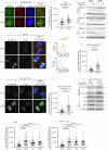
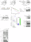
Fig. 1
Fig. 1. PIN1 regulates RNF168 chromatin retention.
A Representative images of RNF168 foci in control (siNTC) or PIN1-depleted cells (siPIN1) after treatment with ionizing radiation (IR), 2 Gy. Scale bars 10 µm. B Quantification of RNF168 foci intensity from A. Data is mean ± s.e.m, n = 170 cells. Source data are provided as a Source Data file. C Western blot of chromatin and soluble fractions or whole cell lysate (WCL) for RNF168, histone H3, vinculin and PIN1. Control and PIN1 depleted U2OS cells were treated with 10 Gy of IR and collected after indicated time points to prepare chromatin, soluble fraction or WCL (performed once). Source data are provided as a Source Data file. D Representative images of myc-RNF168 foci in control (siNTC) or PIN1-depleted cells (siPIN1). Scale bars 10 µm. The right panel shows the fluorescence intensity profiles along the line for myc and γH2AX. E Quantification of myc-RNF168 foci intensity from (D) Data is mean ± s.e.m, n = 113 cells for siNTC and 131 cells for siPIN1. Source data are provided as a Source Data file. F Representative images of myc-RNF168 foci in control (siNTC) or PIN1-depleted cells (siPIN1) after 2 Gy IR. Scale bars 10 µm. G Quantification of myc-RNF168 foci intensity from (F). Data is mean ± s.e.m, n = 142 cells for siNTC and 136 cells for siPIN1. Source data are provided as a Source Data file. H Western blot of chromatin fraction and whole cell lysate (WCL) for myc, histone H3, PIN1 and tubulin in control (siNTC) or PIN1-depleted cells (siPIN1) after treatment with 10 Gy IR. SE: short exposure, LE: Long exposure (Representative of 2 repeats). Source data are provided as a Source Data file. I Quantification of myc-RNF168 intensity after treatment with various PIN1 inhibitors: Juglone (10 µM, 4 hrs), PiB (25 µM, 24 hrs), ATRA (25 µM, 24 hrs). Data is mean ± s.e.m, n = 82 for control, 100 for Juglone and PiB, and 86 for ATRA. Source data are provided as a Source Data file. J Quantification of myc-RNF168 foci intensity after treatment with PIN1 inhibitors and IR. Cells expressing myc-RNF168 were treated with Juglone (10 µM, 4 hrs), PiB (25 µM, 24 hrs), ATRA (25 µM, 24 hrs) before irradiation (2 Gy IR). Data is mean ± s.e.m, n = 82 for control, 84 for Juglone, 77 for PiB, and 74 for ATRA. Source data are provided as a Source Data file.
Fig. 2
Fig. 2. Phosphorylated threonine-208 promotes PIN1 interaction.
A Pull down of endogenous RNF168 by GST-fused WW or W34A mutant domain of PIN1 from U2OS cells (Representative of 2 repeats). Source data are provided as a Source Data file. B Pull down of endogenous RNF168 by GST-fused WW or W34A mutant domain of PIN1 from HeLa cells (Representative of 2 repeats). Source data are provided as a Source Data file. C Pull down of myc-RNF168 by GST-fused WW or W34A mutant domain of PIN1 from U2OS cells treated with 10 Gy of IR. Cell lysates were made at indicated time points after IR (Representative of 2 repeats). Source data are provided as a Source Data file. D Schematic of RNF168 domains and deletion mutant. E Colony survival of U2OS cells depleted of RNF168 and complemented with WT, Δ190-235 RNF168 after treatment with indicated doses of IR. n = 3. Data is mean ± s.e.m. Source data are provided as a Source Data file. F Western blot to show depletion of RNF168 and complementation of RNF168 variants for (E) (performed once). Source data are provided as a Source Data file. G U2OS cells expressing myc-RNF168 wild type (WT) or T208A mutant were subjected to pull down by GST-fused-WW or W34A domain of PIN1 (Representative of 3 repeats). Source data are provided as a Source Data file. H Native mass spectrum of recombinant PIN1 incubated with peptides of the T208 motif from RNF168 at a 1:5 molar ratio (left). Colored peaks indicate PIN1:peptide complex formation between non-phosphorylated peptide (blue) and phosphorylated peptide (green), while black peaks correspond to unbound PIN1. Quantification of the PIN1:peptide peak intensities as a percentage of bound relative to total protein (right). Source data are provided as a Source Data file. I myc-WT-RNF168 or myc-RNF168-T208A were precipitated from HEK293 cells using myc-sepharose beads and probed with anti-myc or anti-phosphorylated-threonine-208 RNF168 (pT208) antibodies (Representative of 2 repeats). Source data are provided as a Source Data file. J HEK293 cells expressing myc-RNF168 were treated with RO-3306 (10 µM) “RO” or roscovitine (25 µM), “Rosc” for 4 hrs and probed with anti- pT208, myc and vinculin antibodies (performed once). Source data are provided as a Source Data file. K Assessment of pT208 and myc following immunoprecipitation of myc-RNF168 from control siRNA-treated cells (siNTC) and from cells treated with siRNAs to the kinases shown (Representative of 2 repeats). Source data are provided as a Source Data file.
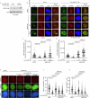
Fig. 3
Fig. 3. P209A mutation rescues T208A-RNF168 hyper-accumulation.
A 1H−1H EXchange SpectroscopY (EXSY) detected activity of SDPV(pT)PK RNF168 peptide with and without PIN1. Left: 2D 1H-1H EXSY spectrum of 2 mM peptide in the absence of PIN1 (mixing time, 300 ms). Middle: 2D 1H-1H EXSY spectrum of 2 mM peptide in the presence of 25 μM PIN1 (mixing time was 300 ms). T and C indicate the diagonal peaks when the proline is in either the trans or cis conformation, respectively. Right: 1H-1H EXSY curves are plotted as the intensity ratio of the C to T peak over the T peak versus mixing times ranging from 12.5 ms to 600 ms and fitted as described in methods. The fitted value of kex is 12.65 ± 0.18 s-1. B Representative images of foci formation of RNF168 mutants. U2OS cells stably expressing siRNA resistant myc-WT-RNF168, T208A, P209A and T208A/P209A mutants. Cells were fixed and stained for myc and γH2AX. Scale bars 10 µm. C As in B, cells expressing myc-WT-RNF168, T208A, P209A, T208A/P209A mutants were treated with 2 Gy of IR and fixed after 1 hr. Cells were stained for myc and γH2AX. Scale bars 10 µm. D Quantification of myc-RNF168 protein foci intensity from (B). Data is mean ± s.e.m, n cells= 73 for WT, 75 for T208A, 92 for P209A and 73 for T208A/P209A. Source data are provided as a Source Data file. E Quantification of myc-RNF168 protein foci intensity from C. Data is mean ± s.e.m, n cells = 152 for WT, 142 for T208A, 130 for P209A and 163 for T208A/P209A. Source data are provided as a Source Data file.
Fig. 4
Fig. 4. PIN1 promotes SUMO2/3ylation of RNF168 and SUMOylation regulates RNF168 chromatin accumulation.
A HEK293 cells complemented with myc-WT-RNF168 or K210R mutant were transfected with 6xHis-Flag-SUMO2. SUMO2 conjugated proteins were enriched by His-Mag Sepharose Ni beads (Ni2+ pull down) under denaturing conditions and detected by western blotting (Representative of 2 repeats). Source data are provided as a Source Data file. B HEK293 cells containing shRNA control (NTC) or shPIN1 were transfected with myc-RNF168 alone or in combination with 6xHis-Flag-SUMO2. Cells were treated with 1 mM IPTG for 72 hrs for PIN1 depletion. SUMO2 conjugated proteins were enriched by His-Mag Sepharose Ni beads (Ni2+ pull down) under denaturing conditions and detected by western blotting (Representative of 2 repeats). Source data are provided as a Source Data file. C HEK293 cells transfected with myc-RNF168-WT, T208A or T208A/P209A along with 6xHis-Flag-SUMO2. SUMO conjugated proteins were pulled down by His-Mag Sepharose Ni beads (Ni2+ pull down) under denaturing conditions. SUMO-conjugated RNF168 variants were detected by western blotting (Representative of 2 repeats). Source data are provided as a Source Data file. D Western blot of SUMO1 and SUMO2/3 conjugates following siRNA treatments (performed once). Source data are provided as a Source Data file. E U2OS cells stably expressing myc-WT-RNF168 were treated with indicated control and SUMO siRNAs, cells were treated with 2 Gy of IR and fixed after 1 hr post IR and stained for myc and γH2AX. Scale bars 10 µm. F Quantification of myc-RNF168 intensity from (E). Data is mean ± s.e.m, n = 90 cells for siNTC, 110 for siSUMO1 and 146 for siSUMO2/3. Source data are provided as a Source Data file. G Western blot of chromatin fraction and WCL for myc, histone H3, SUMO2/3 and tubulin. Control and SUMO2/3 depleted U2OS cells were treated with 10 Gy of IR and collected for fractionation at indicated time points (performed once). Source data are provided as a Source Data file. H Western blot of chromatin fraction and WCL for myc, histone H3 and tubulin. U2OS cells expressing RNF168-WT, T208A or K210R mutants were treated with 10 Gy of IR and collected at indicated time points for fractionation (performed once). Source data are provided as a Source Data file.
Fig. 5
Fig. 5. T208A-RNF168 is resistant to RNF4 and p97/VCP-mediated suppression of accumulation.
A HEK293 cells transfected with GFP-WT-RNF168, GFP-T208A-RNF168 or GFP-K210R-RNF168 and His-myc-Ub. Ubiquitylated proteins were pulled down by His-Mag Sepharose Ni beads (Ni2+ pull down) under denaturing conditions. Ubiquitylated RNF168 was detected by western blotting (Representative of 2 repeats). Source data are provided as a Source Data file. B HEK293 cells were transfected with GFP-WT-RNF168 or GFP-T208A-RNF168, along with HA-RNF4. GFP-Trap precipitation was performed, and precipitated proteins were analyzed using western blotting (Representative of 2 repeats). Source data are provided as a Source Data file. C Quantification of myc-RNF168 foci intensity after radiation. U2OS cells expressing myc-RNF168-WT or myc-RNF168-T208A were treated with siNTC or siRNF4 and stained for myc, 1 hr post IR (2 Gy). myc intensity on y-axis. Data are mean ± s.e.m, n = 153 cells for myc-RNF168-WT+siNTC, 116 for myc-RNF168-WT+siRNF4, 129 for myc-RNF168-T208A+siNTC and 148 for myc-RNF168-T208A+siRNF4. Source data are provided as a Source Data file. D HEK293 cells were transfected with GFP vector alone or with GFP-WT-RNF168. GFP-Trap precipitation was performed, and precipitated proteins were analyzed using western blotting (performed once). Source data are provided as a Source Data file. E Schematic of experimental design for (F). F p97/VCP inhibition results in hyperaccumulation of RNF168 after irradiation. U2OS cells expressing myc-RNF168-WT or T208A mutant were treated with 2 Gy IR and incubated an hour later with 1 μM CB-5083 for another 6 hrs. Cells were stained for myc and γ-H2AX. Scale bars 10 µm (Left). myc intensity on y-axis. Data is mean ± s.e.m, n = 126 cells for myc-RNF168 WT, 108 for myc-RNF168 WT+p97/VCPi, 152 for myc-RNF168 T208A and 159 for myc-RNF168 T208A + p97/VCPi. Source data are provided as a Source Data file.
Fig. 6
Fig. 6. T208A-RNF168 mutant drives increased chromatin ubiquitination, increased 53BP1 accumulation but reduced BRCA1 accumulation.
A Western blot of chromatin fraction for H2AK15ub and H2A from cells expressing siRNA-resistant myc-RNF168-WT or T208A. Endogenous RNF168 is depleted by siRNA. Cells were either left untreated or irradiated by 10 Gy IR and fractionated 1 hr later (Representative of 2 repeats). Source data are provided as a Source Data file. B U2OS cells treated with non-targeting siRNA or RNF168 siRNA and expressing siRNA-resistant myc-WT-RNF168 or myc-T208A-RNF168 were either untreated (left) or treated with 2 Gy of IR (right) and stained for 53BP1 and γH2AX. Scale bars 10 µm. C Quantification of 53BP1foci intensity from untreated cells. Data is mean ± s.e.m, n = 150 for siNTC, 100 for siRNF168, 140 for WT-RNF168 and 124 for T208A-RNF168. Source data are provided as a Source Data file. D Quantification of 53BP1 foci intensity from irradiated cells. Data is mean ± s.e.m, n = 177 for siNTC, 104 for siRNF168, 133 for RNF168-WT and 158 for RNF168-T208A. Source data are provided as a Source Data file. E BRCA1 assessment in U2OS expressing siRNA resistant myc-RNF168-WT, T208A or K210R, depleted of endogenous RNF168 and treated with 2 Gy IR. 2 hrs post IR, cells were stained for BRCA1 and CENPF. Scale bars 10 µm. F Quantification of number BRCA1 foci from (E). Data is mean ± s.e.m, n = 143 cells for siNTC, 153 for siRNF168, 147 for RNF168-WT, 129 for RNF168-T208A and 136 for RNF168-K210R. Source data are provided as a Source Data file. G Quantification of number RAD51 foci in U2OS expressing siRNA-resistant myc-RNF168-WT, T208A or K210R mutants, depleted of endogenous RNF168 and treated with 2 Gy IR. 2 hrs post IR, cells were stained for RAD51 and CENPF (representative images in Supplementary Fig. 6D). Data is mean ± s.e.m, n = 132 cells for siNTC, 207 for siRNF168, 195 for RNF168-WT, 297 for RNF168-T208A and 207 for RNF168-K210R. Source data are provided as a Source Data file.
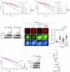
Fig. 7
Fig. 7. The RINF168 SPaCR motif supports radio-resistance through suppression of 53BP1 accumulation.
A Colony survival of U2OS cells depleted of RNF168 and complemented with myc-WT-RNF168, T208A, P209A or T208A/P209A variants after treatment with indicated doses of IR. The number of replicates (n) for each condition is mentioned in parentheses. Data is mean ± s.e.m. p-value compared to siNTC at 2 Gy IR, 7.52 × 10−7 for siRNF168, 0.91 for RNF168-WT, 4.7 × 10−7 for RNF168-T208A, 0.73 for RNF168-P209A and 0.03 for RNF168-T208A/P209A. Source data are provided as a Source Data file. B Colony survival of U2OS cells depleted of RNF168 and complemented with myc-WT-RNF168, T208A, K210R and P209A/K210R variants of myc-RNF168 after treatment with indicated doses of IR. The number of replicates (n) for each condition is mentioned in parentheses. Data is mean ± s.e.m. p-value compared to siNTC at 2 Gy IR, 7.52 × 10−7 for siRNF168, 0.91 for RNF168-WT, 4.7 × 10−7 for RNF168-T208A, 0.00001 for RNF168-K210R and 0.00002 for RNF168-P209A/K210R. Source data are provided as a Source Data file. C Western blot to show depletion of RNF168 and complementation of RNF168 variants for (A) & (B) (performed once). Source data are provided as a Source Data file. D BRCA1 assessment in U2OS expressing siRNA resistant myc-RNF168-WT or T208A, depleted of endogenous RNF168 and 53BP1 and treated with 2 Gy IR. 2 hrs post IR, cells were stained for BRCA1 and CENPF. Scale bars 10 µm. E Quantification of number BRCA1 foci from (D). Data is mean ± s.e.m, n = 77 cells for EV, 82 for myc-RNF168-WT, 92 for RNF168-T208A and 69 for RNF168-T208A+si53BP1. Source data are provided as a Source Data file. F Colony survival of U2OS cells depleted of RNF168 with or without si53BP1 and complemented with myc-WT-RNF168 or T208A after treatment with indicated doses of IR. n = 3. Data is mean ± s.e.m. p-value compared to siNTC at 2 Gy IR, 0.03 for siRNF168, 0.95 for RNF168-WT, 0.0058 for RNF168-T208A and 0.96 for RNF168-T208A + si53BP1. Source data are provided as a Source Data file. G Western blot to show depletion of RNF168, 53BP1 and complementation of RNF168 variants (performed once). Source data are provided as a Source Data file. H Illustration of the post-translational modifications at the RNF168 SPaCR ‘SUMO-PIN1-assisted Chromatin Regulator’ motif to regulate RNF168 dissociation from chromatin.

References

    1. Doil, C. et al. RNF168 binds and amplifies ubiquitin conjugates on damaged chromosomes to allow accumulation of repair proteins. Cell136, 435–446 (2009). - PubMed
    1. Huen, M. S. et al. RNF8 transduces the DNA-damage signal via histone ubiquitylation and checkpoint protein assembly. Cell131, 901–914 (2007). - PMC - PubMed
    1. Kolas, N. K. et al. Orchestration of the DNA-damage response by the RNF8 ubiquitin ligase. Science318, 1637–1640 (2007). - PMC - PubMed
    1. Mailand, N. et al. RNF8 ubiquitylates histones at DNA double-strand breaks and promotes assembly of repair proteins. Cell131, 887–900 (2007). - PubMed
    1. Stewart, G. S. et al. The RIDDLE syndrome protein mediates a ubiquitin-dependent signaling cascade at sites of DNA damage. Cell136, 420–434 (2009). - PubMed

MeSH terms