. 2025 Apr 14;15(1):12849.
doi: 10.1038/s41598-025-97053-9.

A simplified preparation method for single-nucleus RNA-sequencing using long-term frozen brain tumor tissues

Affiliations

A simplified preparation method for single-nucleus RNA-sequencing using long-term frozen brain tumor tissues

Kati J Ernst et al. Sci Rep. .

Abstract

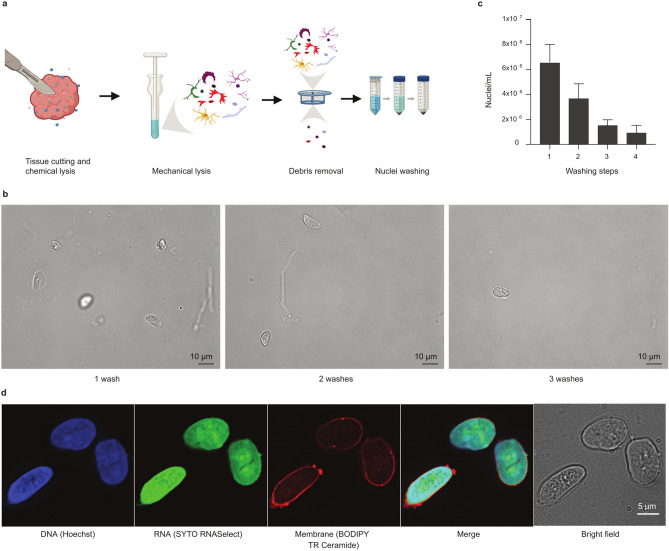
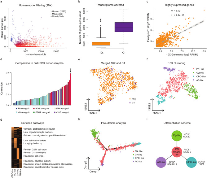
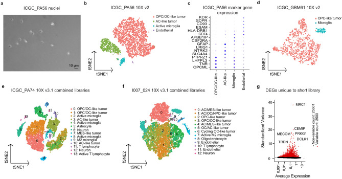
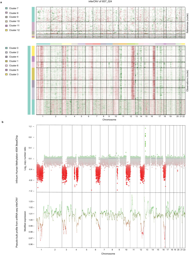
Single-cell RNA-sequencing has provided intriguing new insights into research areas such as developmental processes and tumor heterogeneity. Most approaches, however, rely on the availability of fresh surgical specimens, thereby dramatically reducing the ability to profile particularly rare tissue types. Here, we optimized a method to isolate intact nuclei from long-term frozen pediatric glioma tissues. We performed a technical comparison between different single-nucleus RNA-sequencing (snRNA-seq) systems and applied the established nucleus isolation method to analyze frozen primary glioma tissues. The results show that our fast, simple and low-cost nuclear isolation protocol provides intact nuclei, which can be used in both droplet- and plate-based single-cell sequencing platforms - allowing the identification of distinct tumor cell populations and infiltrating microglia. Additional optimization to include shorter RNA fragments in the 3' sequencing library improved gene detection and cell type annotation. Taken together, the method dramatically increases the potential of studying rare tumor entities and is specifically tailored for using frozen brain tumor tissue.


Conflict of interest statement

Declarations. Competing interests: The authors declare no competing interests.

Figures

Fig. 1
Fig. 1
Isolation of intact nuclei from long-term frozen pediatric glioma tissue. (a) Schematic figure showing sample preparation steps. (b) Representative images showing the effect of an increasing number of washing steps on nuclei yield/integrity and number of debris (scale bar 10 μm). (c) Nuclei yield decreases with increasing number of washes. Data are represented as mean with SEM. (d) Staining for nuclear membrane, DNA and RNA reveals intact nuclei, with no leakage of nucleic acids (scale bars 5 μm). Nuclei originate from a pediatric pilocytic astrocytoma tissue frozen for seven years prior to nuclear extraction. Created with BioRender.com, GraphPad Prism 5 and Affinity Designer 2.
Fig. 2
Fig. 2
Comparison of platforms for studying tumor heterogeneity using patient-derived xenografts. (a) Variance between the numbers of human and mouse transcripts per nuclei from 10X snRNA- seq data of a glioma PDX sample. (b) Number of genes per cell and (c) gene expression levels between 10X and C1 platforms. (d) Comparison to bulk PDX tumor samples. 10X snRNA-seq data of the pHGG PDX sample combined into a pseudo-bulk compared to to bulk microarray data from a group of PDX samples based on correlation. The bulk microarray data of the same sample had the highest correlation to the 10X pseudo-bulk data (marked with an arrow). (e) t-distributed stochastic neighbour embedding (t-SNE) representation of combined 10X and C1 snRNA-seq datasets. (f) t-SNE representation of 10X PDX dataset. (g) Heatmap of pathways enriched among 10X PDX cell types. Colors represent confidence level –log10 (p-val). (h) Pseudotime trajectory analysis of 10X PDX cells. (i) Schematic representation of the identified tumor cell populations. See also Supplementary Fig. S2 and Supplementary Tables S2, S3, S4 and S5 online.
Fig. 3
Fig. 3
SnRNA-seq of fresh frozen primary tumor tissues reveal distinct tumor and healthy cell populations. (a) Brightfield image showing intact isolated nuclei loaded to the 10X system (scale bar 10 μm). (b) t-SNE representation of pilocytic astrocytoma ICGC_PA56 snRNA-seq data. Two tumor clusters, oligodendrocyte precursor/oligodendrocyte -like (OPC/OC-like) and astrocyte-like (AC-like) tumor cells clearly separated from microglia and endothelial cells. (c) The most highly expressed marker genes show differential expression across assigned cell types. (d) OPC-like tumor cells and microglia form separate clusters detected from glioblastoma ICGC_GBM61 snRNA-seq data. 10X v3.1 snRNA-seq data of combined libraries of standard (approx. 300–1000 bp) and short (< 400 bp) cDNA fragments gives a more detailed clustering of the tumors, here (e) pilocytic astrocytoma ICGC_PA74 and (f) pleomorphic xanthoastrocytoma I007_024. (g) Some genes are detected only from the short library data, here ICGC_PA74 as example. See also Supplementary Fig. S3 and S4 and Supplementary Tables S2, S3, S4 and S5 online.
Fig. 4
Fig. 4
Copy number variation analysis supports tumor cell detection from 10 × snRNA- seq data. (a) Copy number variations (CNVs) of single nuclei from 10X snRNA-seq data of I007_024 analyzed by inferCNV. Non-malignant cells are used as control (upper heatmap). (b) A bulk CNV profile of the same tumor derived from Infinium HumanMethylation450 array analysis compared to pseudo-bulk extraction of CNV profiles from 10X data of I007_024 using mean values across the cells. See also Supplementary Fig. S5 and Supplementary Table S4 online.

References

    1. Filbin, M. & Monje, M. Developmental origins and emerging therapeutic opportunities for childhood cancer. Nat. Med.25, 367–376. 10.1038/s41591-019-0383-9 (2019). - PMC - PubMed
    1. Sturm, D., Pfister, S. M. & Jones, D. T. W. Pediatric gliomas: Current concepts on diagnosis, biology, and clinical management. J. Clin. Oncol.35, 2370–2377. 10.1200/JCO.2017.73.0242 (2017). - PubMed
    1. Liu, K. W., Pajtler, K. W., Worst, B. C., Pfister, S. M. & Wechsler-Reya, R. J. Molecular mechanisms and therapeutic targets in pediatric brain tumors. Sci. Signal10.1126/scisignal.aaf7593 (2017). - PubMed
    1. Kallappagoudar, S., Yadav, R. K., Lowe, B. R. & Partridge, J. F. Histone H3 mutations–a special role for H3.3 in tumorigenesis?. Chromosoma124, 177–189. 10.1007/s00412-015-0510-4 (2015). - PMC - PubMed
    1. Danilenko, M. et al. Single-cell DNA sequencing identifies risk-associated clonal complexity and evolutionary trajectories in childhood medulloblastoma development. Acta Neuropathol.144, 565–578. 10.1007/s00401-022-02464-x (2022). - PMC - PubMed

LinkOut - more resources