. 2025 Apr 14;15(1):12774.
doi: 10.1038/s41598-025-96136-x.

Escherichia coli type I toxin TisB exclusively controls proton depolarization following antibiotic induced DNA damage

Affiliations

Escherichia coli type I toxin TisB exclusively controls proton depolarization following antibiotic induced DNA damage

Tekle Airgecho Lobie et al. Sci Rep. .

Abstract

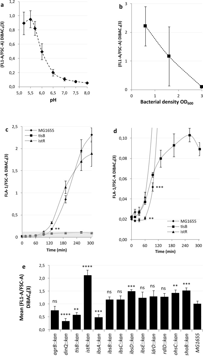
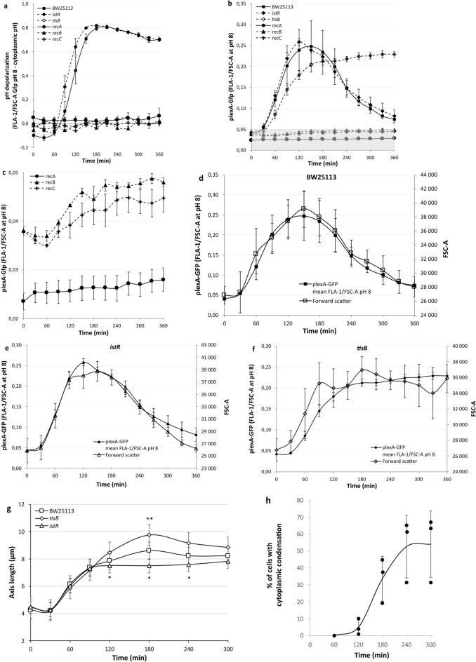
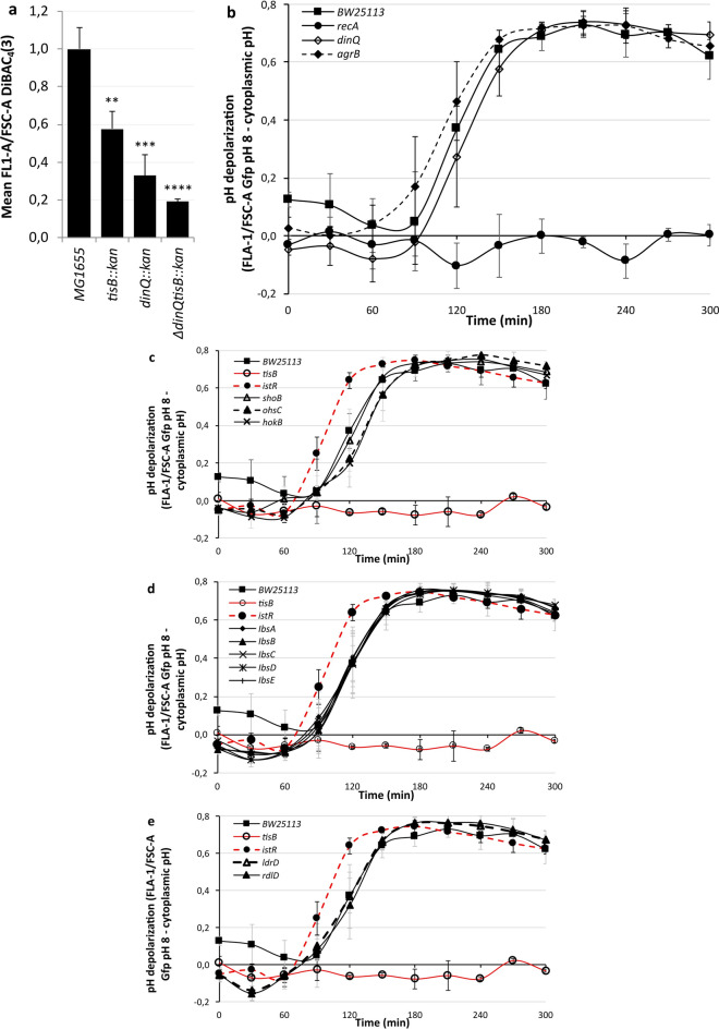
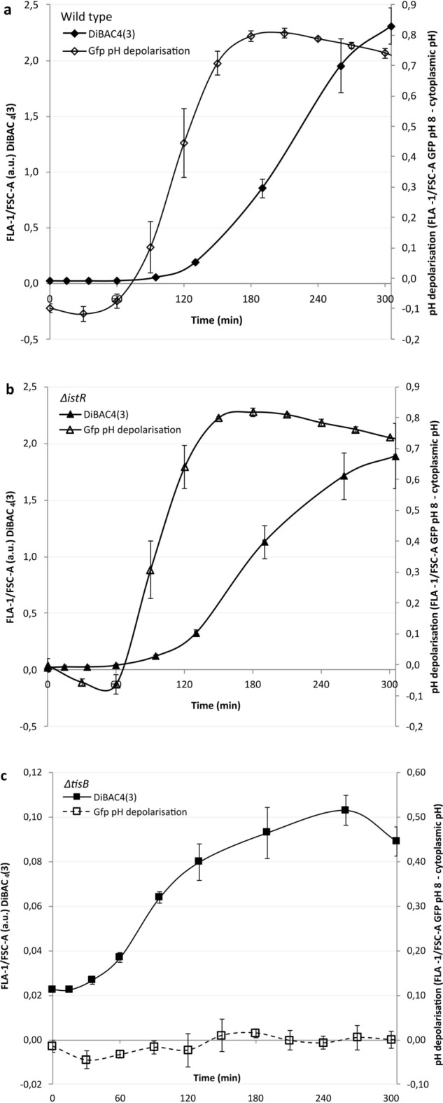
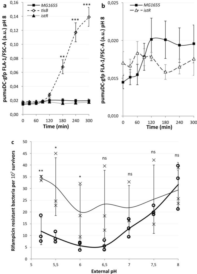
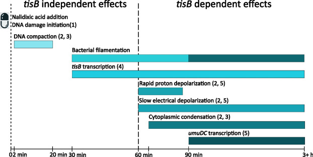
Bacterial toxin-antitoxin (TA) systems are genetic loci where the antitoxin gene product helps to control the expression or activity of the toxin gene product. Type I TA systems typically produce hydrophobic peptides that often localize to the inner membrane of bacteria. These amphipathic peptides can then potentially affect ion flows across the inner membrane. Here, we show that several type I toxins from Escherichia coli can affect depolarization, whereas tisB exclusively controls the depolarization of the proton gradient. tisB has been linked to persister cell formation following treatment with the antibiotic ciprofloxacin and tisB-istR has been implicated in the control of proton depolarization following treatment with ofloxacin. These results suggest that tisB could initiate the formation of persister cells by fully dissipating the proton gradient and that most of the electrical gradient greatly limiting ATP production following antibiotic-induced DNA damage.

Keywords: E. coli; Antibiotics; DNA damage; Membrane depolarization; TisB; Type I toxin antitoxin systems.


Conflict of interest statement

Delcaration. Competing interests: The authors declare that they have no competing interests.

Figures

Fig. 1
Fig. 1
Multiple type I TA systems can result in loss of electrical potential following antibiotic-induced DNA damage. a DiBAC4(3) is most sensitive to the electrical potential at pH values of 5.2–5.7. Bacteria were grown to the exponential phase in LBK at the pH shown before being treated with 100 µg/ml nalidixic acid. The samples were further incubated for five hours before being stained with DiBAC4(3) in PBS buffered to the same pH as that of the media. b DiBAC4(3) is most sensitive at low bacterial densities. Bacteria were grown to the exponential phase in LBK at pH 5.7 before being treated with 100 µg/ml nalidixic acid. The samples were further incubated for five hours before staining with DiBAC4(3) in PBS, pH 5.7. c-d tisB and istR begin electrical depolarization before the wild type but achieve lower total signals after five hours, with tisB achieving little total depolarization. Bacteria were grown to OD600 = 0.6 in LBK at pH 5.7 before being treated with 100 µg/ml nalidixic acid. The samples were taken at the time points indicated and stained with DiBAC4(3) in PBS, pH 5.7. The gray shaded area in c illustrates the region of the diagram examined in d. The statistical significance refers to MG1655 vs istR. Significance for tisB vs istR was reached after 95 min and was at least p = 0.008 (Supplementary data file). Significance was calculated using a two tailed unpaired T-test. e Multiple type I TA mutants can result in reduced electrical depolarization. Bacteria were grown to OD600 = 0.6 in LBK at pH 5.7 before being treated with 100 µg/ml nalidixic acid. The samples were removed after five hours and stained with DiBAC4(3) in PBS, pH 5.7. All data points shown are means ± standard deviations, n = 3 or 4 (independent biological replicates). Statistical significance was assessed using a one-way ANOVA followed by Dunnett’s post hoc test for multiple comparisons (Supplementary data file).
Fig. 2
Fig. 2
Induction of tisB expression affects the extent of pH depolarization and flow cytometry scattering. All strains (a-f) contained plexA-gfp and were incubated at pH 5.7 in LBK. Nalidixic acid (100 µg/ml) was added at zero minutes, resulting in immediate translation of pH-sensitive Gfp. a At the indicated time points, the fluorescence density of the bacteria was recorded at cytoplasmic pH and at pH 8 by depolarising the bacteria with sodium benzoate. The difference in fluorescence intensity was corrected for the amount of Gfp present as measured by fluorescence density at pH 8. No statistical significance was seen between BW25113 and istR using a two tailed, unpaired T-test. BW25113 was statistically significant from tisB at 120 min. (Supplementary data file). b At the indicated time points, the fluorescence density of the bacteria was recorded at pH 8 with the use of sodium benzoate. This corresponds to the activity of the lexA promoter. The gray shaded area in b illustrates the region of the diagram examined in c. c As with b but focusing on the reduced signals from recA-C. The difference between recA and recB or recC was statistically significant at all time points. No difference was seen between recB and recC using a two tailed, unpaired T-test (Supplementary data file). Increases in signal intensity were significant between 60 min, the low, and 360 min for the recB and recC mutants, p = 0.0016 and 0.0319 respectively. d-f Comparison of lexA promoter activity and the extent of forward scattering (FSC-A). g Comparison of the extent of filamentation as determined by microscopy. Statistical significance between istR and tisB was achieved from 120–240 min and significance between BW25113 and tisB at 180 min using a two tailed, unpaired T-test (Supplementary data file). h Kinetics of cytoplasmic condensation in wild type bacteria at pH 5, individual data points and the mean are shown. Time is the time since the addition of nalidixic acid. All data points shown are means ± standard deviations, n = 3 (n =  > 53 in g, n = 100–300 in h) (independent biological replicates were obtained).
Fig. 3
Fig. 3
tisB is responsible for pH depolarization. In all cases, the strains were grown to the exponential phase in LBK before being exposed to 100 µg/ml nalidixic acid. a A double dinQ tisB mutant results in significantly less DiBAC4(3) staining than either dinQ or tisB alone following antibiotic-induced DNA damage. The fluorescence density measurements of bacteria stained with DiBAC4(3) was measured on a flow cytometer after five hours of incubation with nalidixic acid. A two tailed, unpaired T-test was used to assess significance (Supplementary data file). b-e Differences in the fluorescence density measurements of bacteria containing Gfp at the intracellular pH and at pH 8, through the use of the weak acid sodium benzoate, were calculated at the time points indicated. b dinQ and agrB mutants varied only slightly from the wild type rate of proton gradient dissipation whilst recA mutants did not equilibrate. c tisB mutants did not dissipate the proton gradient, whereas istR mutants presented an accelerated rate of dissipation compared with the wild-type. Compared with those of the wild type, the proton gradient dissipation rates of the shoB, ohsC and hokB mutants did not differ. d None of the ibsA-E mutants deviated from the wild-type rate of proton gradient dissipation. e Neither ldrD nor the rdlD mutants showed any difference in rate from the wild-type pH equilibration rate. All data points shown are means ± standard deviations, n = 4 a, 5 b-e (independent biological replicates).
Fig. 4
Fig. 4
The pH gradient dissipates faster than the electrical potential. In all cases, exponentially growing strains in LBK buffered to pH 5.7 were exposed to 100 µg/ml nalidixic acid at time = zero minutes. Fluorescence density measurements of either DiBAC4(3) to measure the electrical potential (closed symbols) or Gfp to measure the pH gradient (open symbols) were monitored using flow cytometry at the time points indicated. The pH gradient is the difference in fluorescence intensity at the intracellular pH versus that at pH 8 divided by the amount of Gfp present. a Wild-type E. coli. b istR deletion mutants show faster depolarization of both potentials as compared to the wild type and c tisB deletion mutants show no proton depolarization but fast initial electrical depolarization compared with the wild type, which fails to accelerate after 60 min. All data points shown are the means ± standard deviations, n = 4 or 5 (independent biological replicates).
Fig. 5
Fig. 5
Failure of tisB mutants to depolarize results in elevated umuDC promoter activity and mutagenesis in low pH environments. The rate of proton depolarization in low-pH environments determines the extent to which umuDC transcription is inhibited. a The tisB mutant, which cannot depolarise the proton gradient, results in large increases in umuDC compared with the background strains. Flow cytometry measurements are of the GFP fluorescence density of a umuDC promoter GFP construct, over time. Exponentially growing cultures at pH 5.7 were treated at 0 min with 100 ug/ml nalidixic acid to induce the SOS response. All fluorescence readings were quantified at pH 8. A two tailed, unpaired T-test was used to assess significance (Supplementary data file). b The istR mutant, with an accelerated rate of depolarization, led to no increase in fluorescence density, whereas the wild type presented a small but not significant increase (Supplementary data file). c Increased mutagenesis is observed in the tisB mutant, a cross marker, at low external pH compared with the wild type, open circle marker. Exponentially growing cultures were exposed to UV light (20 J) before further growth and subsequent plating on rifampicin plates (100 µg/ml). A two tailed, unpaired T-test was used to assess significance (Supplementary data file). All data points shown are means ± standard deviations, n = 3 or 4 (independent biological replicates).
Fig. 6
Fig. 6
TisB dependent and Associated Cellular Events Following DNA Damage in E. coli. The addition of nalidixic acid, a DNA-damaging antibiotic, triggers a cascade of cellular events through inhibition of DNA gyrase and topoisomerase IV. This inhibition leads to blocked replication and transcription which ultimately results in DNA double-strand breaks. These breaks are processed by RecBCD, leading to RecA loading onto ssDNA. The resulting nucleoprotein filament catalyzes LexA autodegradation, activating the SOS response. Cellular responses occur in a defined temporal sequence: DNA compaction initiates within minutes and peaks at 20 min post-treatment,. Cell filamentation, driven by SulA-mediated FtsZ inhibition, begins at 30 min and continues to increase for up to 3 h (Fig. 2g). tisB transcription begins to increase after 30 min. TisB-dependent effects emerge at 60–90 min post-treatment, coinciding with membrane depolarization (disruption of electrical (Fig. 1c) and proton gradients (Fig. 2a),) and subsequent cytoplasmic condensation (Fig. 2h). Filamentation becomes tisB dependent at this stage (Fig, 2g). At later stages the umuDC promoter transcription levels are highly tisB dependent (Fig. 5a). These events can lead to bacterial persistence under specific conditions,. While the presented timings are indicative and illustrative, the relative ordering of events is expected to be maintained. Observed timings will vary depending on environmental conditions and whether bulk or single-cell readings are taken.

Similar articles

References

    1. Yamaguchi, Y. & Inouye, M. Regulation of growth and death in escherichia coli by toxin-antitoxin systems. Nat. Rev. Microbiol.9, 779–790. 10.1038/nrmicro2651 (2011). - PubMed
    1. Darfeuille, F., Unoson, C., Vogel, J. & Wagner, E. G. H. An antisense RNA inhibits translation by competing with standby ribosomes. Mol. Cell26, 381–392. 10.1016/j.molcel.2007.04.003 (2007). - PubMed
    1. Kristiansen, K. I., Weel-Sneve, R., Booth, J. A. & Bjoras, M. Mutually exclusive RNA secondary structures regulate translation initiation of DinQ in escherichia coli. RNA22, 1739–1749. 10.1261/rna.058461.116 (2016). - PMC - PubMed
    1. Weel-Sneve, R. et al. Single transmembrane peptide DinQ modulates membrane-dependent activities. PLoS Genet.9, e1003260. 10.1371/journal.pgen.1003260 (2013). - PMC - PubMed
    1. Kawano, M., Oshima, T., Kasai, H. & Mori, H. Molecular characterization of long direct repeat (LDR) sequences expressing a stable mRNA encoding for a 35-amino-acid cell-killing peptide and a cis-encoded small antisense RNA in escherichia coli. Mol. Microbiol.45, 333–349. 10.1046/j.1365-2958.2002.03042.x (2002). - PubMed

MeSH terms

LinkOut - more resources