. 2025 Aug;62(8):10652-10669.
doi: 10.1007/s12035-025-04864-y. Epub 2025 Apr 14.

Remote Ischemic Postconditioning Improve Cerebral Ischemia-Reperfusion Injury Induced Cognitive Dysfunction through Suppressing Mitochondrial Apoptosis in Hippocampus via TK/BK/B2R-Mediated PI3K/AKT

Affiliations

Remote Ischemic Postconditioning Improve Cerebral Ischemia-Reperfusion Injury Induced Cognitive Dysfunction through Suppressing Mitochondrial Apoptosis in Hippocampus via TK/BK/B2R-Mediated PI3K/AKT

Haocheng Qin et al. Mol Neurobiol. 2025 Aug.

Abstract

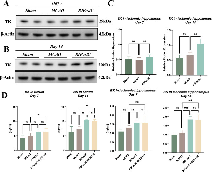
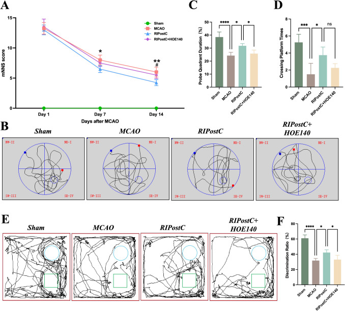
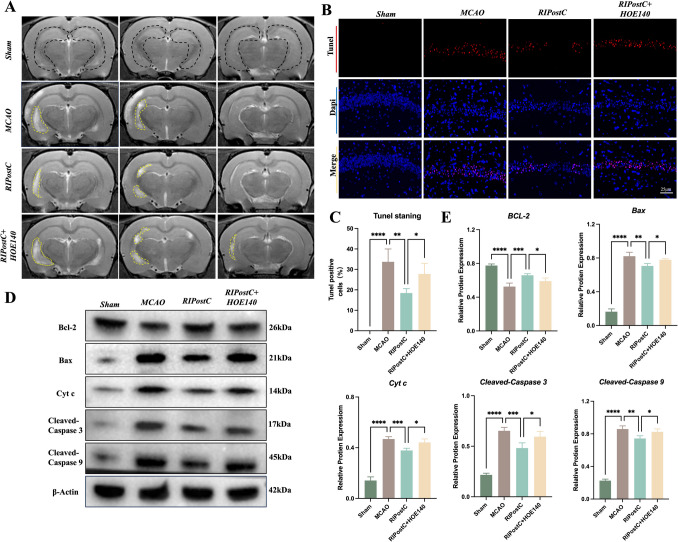
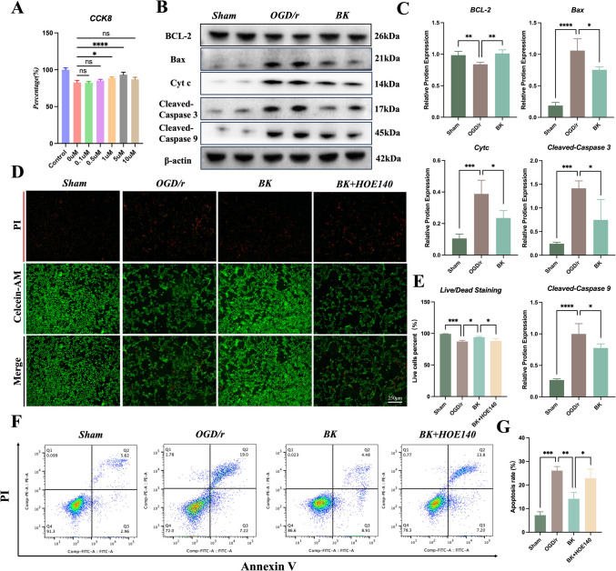
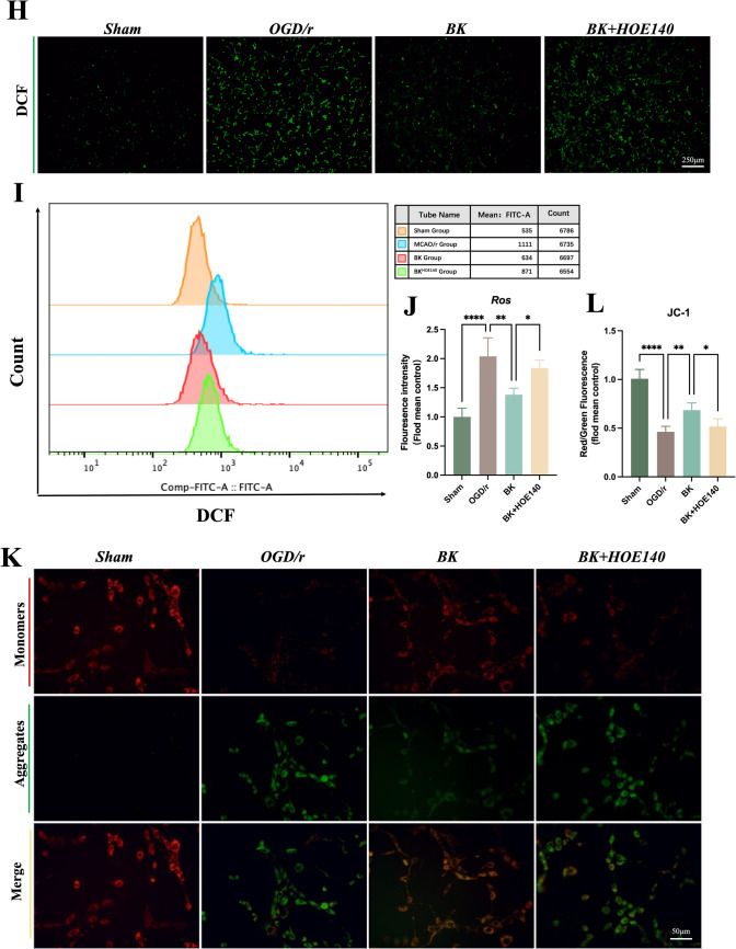
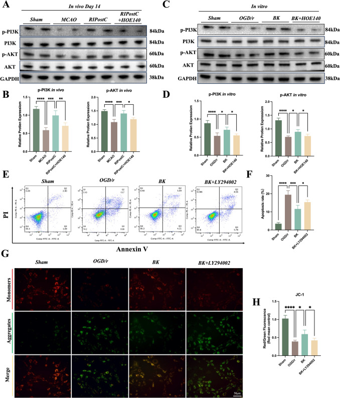
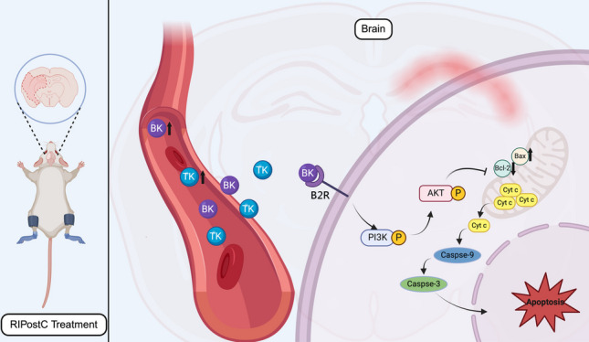
Remote ischemic postconditioning (RIPostC) is known to improve motor function recovery in animal models, but its efficacy in alleviating cognitive impairment caused by ischemic stroke remains unclear. This study aims to investigate the beneficial role of RIPostC in recovering cognitive impairment induced by cerebral ischemia-reperfusion injury (CIRI). Building upon our previous research findings, we proved that the TK/BK/B2R pathway is crucial for understanding the crosstalk between cognitive impairment and RIPostC. Additionally, in vitro experiments were conducted using the oxygen glucose deprivation/re-oxygenation (OGD/r) HT-22 cell model, which revealed that the mechanism by which RIPostC suppressed mitochondrial apoptosis was mainly through the activation of the B2R/PI3K/AKT signaling pathway, thereby protecting neurons in the ischemic hippocampus from ischemic damage. To investigate the effect of RIPostC on cognitive function recovery following ischemic stroke, we established a rat model using left middle cerebral artery occlusion reperfusion (MCAO/r). 48 h after MCAO/r, rats were subjected to 3 circles of RIPostC therapy daily for 12 consecutive days. HOE140 was used to antagonize the bradykinin 2 receptor (B2R). Cognitive function was assessed using a modified neurological severity score, the Morris water maze, and the novel object recognition test. Local infarct volume in the hippocampus was measured through MRI scanning. The apoptosis rate of hippocampal neurons was quantified using TUNEL staining. Protein expression levels of kallikrein (TK) and mitochondrial apoptosis-related proteins, Cyt c, Bcl-2, Bax, cleaved caspase-3, and cleaved caspase-9, were detected in ischemic hippocampal tissue using Western blot (WB). The expression of bradykinin (BK) in serum and the ischemic penumbra was measured using an enzyme-linked immunosorbent (ELISA) assay. In the cell experiments, the HT-22 cell line and OGD/r model were used to simulate in vitro hippocampal ischemia. WB was performed to detect the expression of apoptosis-related proteins and PI3K/AKT pathway proteins. The apoptosis rate of HT-22 cells was detected using Annexin-V/PI flow cytometry and a cell viability kit. JC-1 staining and reactive oxygen species staining were used to evaluate mitochondrial condition. The PI3K/AKT pathway was inhibited using LY294002. RIPostC significantly upregulated the concentrations of TK and BK in the ischemic hippocampus. Behavioral function tests demonstrated that daily RIPostC therapy for 12 days significantly promoted cognitive function recovery in MCAO/r rats. Through MRI analysis, we found that RIPostC therapy effectively reduced the infarct volume in the hippocampus. Additionally, TUNEL staining and WB results of apoptosis-related proteins showed that RIPostC therapy significantly reduced apoptosis of hippocampal neurons. However, the therapeutic effect of RIPostC was reversed by the B2R antagonist HOE14, indicating that the TK/BK/B2R pathway mediated the neuroprotective effect of RIPostC. Cell experiments further confirmed that BK/B2R significantly attenuated mitochondrial apoptosis induced by ischemia-hypoxia injury in HT-22 cells. In vivo and in vitro results from WB demonstrated that the BK/B2R pathway activated the PI3K/AKT signaling pathway. Finally, the PI3K inhibitor LY294002 reversed the anti-apoptotic effect induced by BK/B2R. RIPostC therapy effectively inhibited mitochondrial apoptosis of hippocampal neurons and significantly alleviated cognitive dysfunction associated with CIRI by regulating the TK/BK/B2R-medated PI3K/AKT pathway. In conclusion, RIPostC represents a promising therapeutic strategy for combating cognitive dysfunction by inhibiting cell apoptosis in hippocampus. Moreover, our results suggest that RIPostC may have a broader protective effect against apoptosis in other ischemia-reperfusion-related diseases.

Keywords: Apoptosis; Cerebral ischemia–reperfusion injury; Cognitive dysfunction; Hippocampus; Remote ischemic postconditioning.


Conflict of interest statement

Declarations. Competing Interests: The authors declare no competing interests.

Figures

Fig. 1
Fig. 1
Experimental design and Laser speckle contrast analysis images of rat paws. (A) Experimental design for animal experiments. (B) Experimental design for cell experiment. (C) Laser speckle blood flow images of rat paws before RIPostC intervention, during the intervention and after blood perfusion
Fig. 2
Fig. 2
RIPostC increase TK expression in ischemic hippocampus on day 14 and increase BK expression in serum and ischemic hippocampus on day 14. The protein concentration of TK in ischemic hippocampus was evaluated through Western blot. The expression of BK in ischemic hippocampus and serum was evaluated through ELSIA (A-B) Representative Western blot grave value image of TK in ischemic hippocampus on day 7 and day 14 respectively. (C) Protein concentration was normalized to β-Actin and statically analyzed among four groups (n = 4). (D) The concentration of BK in serum and ischemic hippocampus were measured by ELISA assay (n = 4). * p < 0.05, **p < 0.01
Fig. 3
Fig. 3
The beneficial effect of RIPostC on cognitive behavioral function through activating B2R. On the 14th day after MCAO/r, cognition-related behaviors of rats in Sham group, MCAO group, RIPostC group and RIPostC + HOE140 group were evaluated via mNSS, MWM and NORT analysis. The mNSS, probe quadrant duration, platform crossings times and discrimination ration were recorded and analyzed. (A) mNSS among different groups was statically analyzed (n = 4), *p < 0.05, **p < 0.01 RIPostC compared to MCAO. # p < 0.05, RIPostC + HOE140 compared to RIPostC. (B-D) Probe quadrant duration and platform crossings times among different groups was statically analyzed (n = 4), ns > 0.05, *p < 0.05, ***p < 0.001, ****p < 0.0001. (E–F) The discrimination rate (calculated as N/(N + F) × 100%) among different groups was statically analyzed (n = 4), *p < 0.05, ****p < 0.0001
Fig. 4
Fig. 4
RIPostC decreased infarct size and neurons apoptosis in hippocampus via B2R. On the 14th day after MCAO/r, infarct size, apoptosis rate of neurons, and mitochondria apoptosis-related protein in hippocampus Sham group, MCAO group, RIPostC group and RIPostC + HOE140 group were evaluated. (A) The Hippocampus regions were highlighted by black frame in Sham group. Infarct size in hippocampus were highlighted by yellow frame in other groups. T2 high signal representing ischemic region of coronal plane in hippocampus (× 40 objective). (B) Representative Tunel staining of apoptosis neuron in CA1 region of ischemic hippocampus on day 14. (× 40 objective). (C) Tunel positive neurons among different groups was counted and statically compared (n = 4). * p < 0.05, ** p < 0.01, ***p < 0.001, ****p < 0.0001. Bar = 200 μm. (D) Representative Western blot grave value image of Bcl-2, Bax, Cyt c, Cleaved-Caspase 3 and Cleaved-Caspase 9 in ischemic hippocampus on day14. (E) Protein concentration was normalized to β-Actin and statically analyzed among four groups (n = 4). * p < 0.05, ** p < 0.01, ***p < 0.001, ****p < 0.0001
Fig. 5
Fig. 5
BK significantly reduced OGD/r-mediated apoptosis of HT-22 cells through activating B2R. OGD/r model of HT-22 cells was used to simulate ischemic hippocampus in vivo. Western Blot, Calcein AM/PI staining, ROS detection, flow cytometry of apoptosis and JC-1 staining were used to evaluate the changes of apoptosis ratio and mitochondria function among four groups. (A) CCK8 assay for determining most suitable concentration of BK. Group treated with different concentration were all normalized to Control group (n = 4). *p < 0.05, ****p < 0.0001, n = 4. (B) Representative Western blot grave value image of Bcl-2, Bax, Cyt c, Cleaved-Caspase 3 and Cleaved-Caspase 9 in HT-22 from different groups. (C) Protein concentration was normalized to β-Actin and statically analyzed among four groups (n = 4). * p < 0.05, ** p < 0.01, ***p < 0.001, ****p < 0.0001. (D) Representative images of Calcein AM/PI staining for live (labeled with green fluorescence) or dead cells (labeled with red fluorescence) (× 40 objective). (E) Proportion of live cells was counted through image J and statically analyzed among four groups (n = 4), * p < 0.05, ***p < 0.001. (F) Representative images of flow cytometry for apoptotic cells. In the upper right quadrant are the late modulated cells, while lower right quadrant are the early modulated cells. (G) Apoptotic cells among four groups were statically analyzed (n = 4), * p < 0.05, ** p < 0.01, ***p < 0.001. (H) Representative images of ROS production in HT-22 cells, bar = 1 mm.(I) ROS production in HT-22 cells among four group was evaluated through flow cytometry. (J) Fluorescence was calculated with image J and statically analyzed among four groups (n = 4), * p < 0.05, ** p < 0.01, ***p < 0.001. (× 10 objective) (K) Representative images of JC-1 staining for MMP. Green labels disappear of MMP, Red label normal MMP. (L) Ratio of Red and Green fluorescence was calculated and normalized to Sham group. Fold mean control was statistically analyzed. * p < 0.05, ** p < 0.01, ***p < 0.001. (× 40 objective)
Fig. 5
Fig. 5
BK significantly reduced OGD/r-mediated apoptosis of HT-22 cells through activating B2R. OGD/r model of HT-22 cells was used to simulate ischemic hippocampus in vivo. Western Blot, Calcein AM/PI staining, ROS detection, flow cytometry of apoptosis and JC-1 staining were used to evaluate the changes of apoptosis ratio and mitochondria function among four groups. (A) CCK8 assay for determining most suitable concentration of BK. Group treated with different concentration were all normalized to Control group (n = 4). *p < 0.05, ****p < 0.0001, n = 4. (B) Representative Western blot grave value image of Bcl-2, Bax, Cyt c, Cleaved-Caspase 3 and Cleaved-Caspase 9 in HT-22 from different groups. (C) Protein concentration was normalized to β-Actin and statically analyzed among four groups (n = 4). * p < 0.05, ** p < 0.01, ***p < 0.001, ****p < 0.0001. (D) Representative images of Calcein AM/PI staining for live (labeled with green fluorescence) or dead cells (labeled with red fluorescence) (× 40 objective). (E) Proportion of live cells was counted through image J and statically analyzed among four groups (n = 4), * p < 0.05, ***p < 0.001. (F) Representative images of flow cytometry for apoptotic cells. In the upper right quadrant are the late modulated cells, while lower right quadrant are the early modulated cells. (G) Apoptotic cells among four groups were statically analyzed (n = 4), * p < 0.05, ** p < 0.01, ***p < 0.001. (H) Representative images of ROS production in HT-22 cells, bar = 1 mm.(I) ROS production in HT-22 cells among four group was evaluated through flow cytometry. (J) Fluorescence was calculated with image J and statically analyzed among four groups (n = 4), * p < 0.05, ** p < 0.01, ***p < 0.001. (× 10 objective) (K) Representative images of JC-1 staining for MMP. Green labels disappear of MMP, Red label normal MMP. (L) Ratio of Red and Green fluorescence was calculated and normalized to Sham group. Fold mean control was statistically analyzed. * p < 0.05, ** p < 0.01, ***p < 0.001. (× 40 objective)
Fig. 6
Fig. 6
RIPostC reduced ischemia-hypoxia-mediated mitochondrial apoptosis of hippocampus neurons through activating PI3K/AKT intracellular pathway. Protein expression of p-PI3K/PI3K and p-AKT/ATK was detected in both MCAO/r and OGD/r model. LY294002 was used to inhibit phosphorylation of PI3K and confirm the potential interaction between BK and PI3K/AKT pathway through flow cytometry and JC-1 staining (A) Representative Western blot grave value image of differences of PI3K/AKT pathway in rats from different groups (n = 4). (B) Protein concentration was normalized to GAPDH and statically analyzed among four groups (n = 4). * p < 0.05, ****p < 0.0001. (C) Representative Western blot grave value image of differences of PI3K/AKT pathway in HT-22 from different groups (n = 4). (B, D) Protein concentration was normalized to GAPDH and statically analyzed among four groups (n = 4), * p < 0.05, ** p < 0.01, ***p < 0.001, ****p < 0.0001. (E) Representative images of flow cytometry of apoptotic HT-22 cells. In the upper right quadrant are the late modulated cells, while lower right quadrant are the early modulated cells. (F) Apoptotic cells among four groups were statically analyzed (n = 4), * p < 0.05, ***p < 0.001. (G) Representative images of JC-1 staining for MMP. Green labels disappear of MMP, Red label normal MMP, (× 20 objective). (H) Ratio of Red and Green fluorescence was calculated and normalized to Sham group. Fold mean control was statistically analyzed. * p < 0.05, ***p < 0.001
Fig. 7
Fig. 7
Schematic depiction of this research

Similar articles

References

    1. Hankey GJ (2017) Stroke. Lancet 389(10069):641–654 - PubMed
    1. Rundek T et al (2022) Vascular Cognitive Impairment (VCI). Neurotherapeutics 19(1):68–88 - PMC - PubMed
    1. Rost NS et al (2022) Post-stroke cognitive impairment and dementia. Circ Res 130(8):1252–1271 - PubMed
    1. Huang YY et al (2022) Post-stroke cognitive impairment: epidemiology, risk factors, and management. J Alzheimers Dis 86(3):983–999 - PubMed
    1. Gibson E et al (2022) Occupational therapy for cognitive impairment in stroke patients. Cochrane Database Syst Rev 3(3):Cd006430 - PMC - PubMed

MeSH terms

Substances

LinkOut - more resources