. 2025 Sep;46(9):2496-2508.
doi: 10.1038/s41401-025-01553-3. Epub 2025 Apr 14.

Lipin1-dependent transcriptional inactivation of SREBPs contributes to selinexor sensitivity in multiple myeloma

Affiliations

Lipin1-dependent transcriptional inactivation of SREBPs contributes to selinexor sensitivity in multiple myeloma

Jun-Ying Wang et al. Acta Pharmacol Sin. 2025 Sep.

Abstract

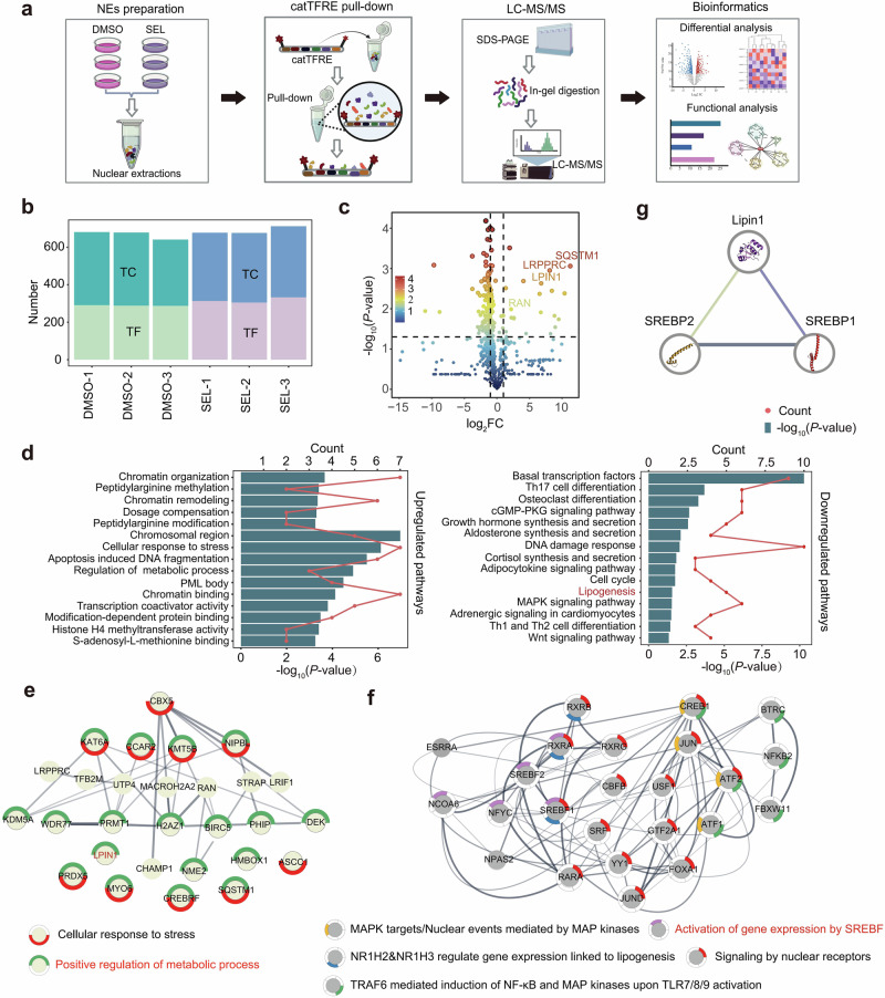
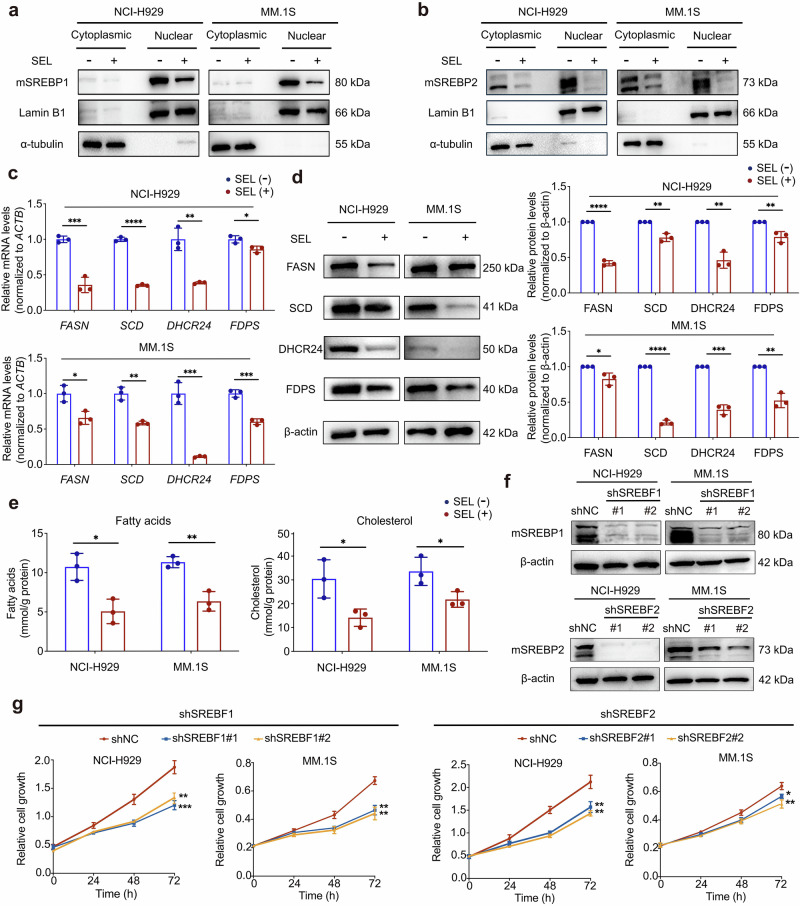
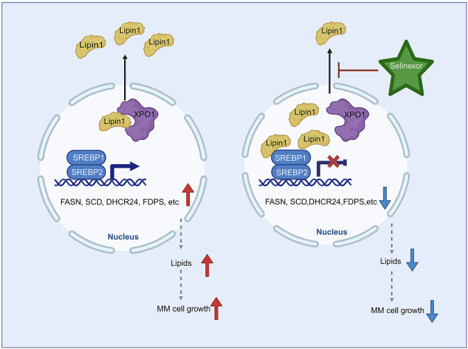
Selective nuclear export inhibitor selinexor (SEL) represents a promising therapeutic strategy for relapsed/refractory multiple myeloma (RRMM). But its mechanisms of action as well as factors that influence therapeutic responses have not been fully characterized yet. In this study we employed catTFRE proteomics technique to profile changes in nuclear abundance of activated transcription factors (TFs)/co-factors (TCs) in myeloma cells following SEL treatment. We found that pharmacological inhibition of exportin-1 (XPO1) by SEL leads to a significant nuclear accumulation of Lipin1 in NCI-H929 cells. Nuclear-localized Lipin1 acted as a transcriptional cofactor that suppressed the transcriptional activity of SREBPs. By performing subcellular localization analysis, molecular docking, co-immunoprecipitation and other assays, we demonstrated that Lipin1 was subjected to XPO1-dependent nuclear export. We demonstrated that SEL downregulated the expression of key lipogenesis-related genes regulated by SREBPs including FASN, SCD, DHCR24 and FDPS, leading to reduced fatty acid and cholesterol synthesis in MM cell lines and primary CD138+ cells. Using shRNA-mediated knockdown assays, we elucidated the critical role of Lipin1 in mediating the inhibitory effects of SEL on the SREBPs pathway and its contribution to SEL sensitivity both in vitro and in murine xenograft models. In conclusion, we reveal a novel mechanism by which SEL downregulates cellular lipid biosynthesis, thereby inhibiting the proliferation of myeloma cells. This study highlights the critical role of Lipin1 in the anti-myeloma effects of SEL, suggesting its potential as a biomarker for identifying patients who are most likely to benefit from SEL-based therapies.

Keywords: Lipin1; SREBPs; exportin-1; multiple myeloma; proteomics.; selinexor.


Conflict of interest statement

Competing interests: The authors declare no competing interests.

Figures

Fig. 1
Fig. 1. catTFRE proteomics analysis revealed SEL-induced nuclear accumulation of TC molecule Lipin1 in MM cells.
a Schematic illustration of the experimental procedure for catTFRE proteomics detection. NCI-H929 cells were treated with either DMSO (n = 3) or 500 nM SEL (n = 3) for 12 h followed by nuclear protein extraction, catTFRE pull-down, mass spectrometry detection and bioinformatics analysis. b The bar plot indicated the number of TFs and TCs detected in each sample. c The volcano plot illustrates the significantly altered TFs/TCs in SEL-treatment group compared to DMSO-treatment group. d Pathway enrichment analysis of upregulated and downregulated TFs/TCs in MM nucleus following SEL treatment. e, f Molecular network of upregulated (e) or downregulated (f) TF/TCs, along with the biological processes in which they are involved. g Protein-protein interaction analysis revealed that Lipin1 interacts with SREBP1 and SREBP2.
Fig. 2
Fig. 2. Lipin1 is subjected to XPO1-dependent nuclear export and can be retained in the nucleus by inhibiting XPO1.
a Immunofluorescence analysis of the subcellular localization of Lipin1 (green) in NCI-H929 and MM.1S following DMSO or 500 nM SEL treatment for 12 h. Cell nuclei were stained with DAPI (blue). Scale bar: 20 μm. b Western blot analysis of Lipin1 levels in cytoplasmic and nuclear extractions from MM cell lines treated with DMSO or 500 nM SEL for 12 h. Lamin B1 was used as a loading control for nuclear protein, α-tubulin served as a loading control for cytoplasmic protein. c Western blot analysis of Lipin1 levels in cytoplasmic and nuclear extractions from MM cell lines with or without XPO1 knockdown. Lamin B1 was used as a loading control for nuclear protein, α-tubulin served as a loading control for cytoplasmic protein. d The molecular docking analysis revealed an interaction between XPO1-RanGTP (upper panel, colored in blue) and Lipin1 (lower panel, colored in green). e Co-IP assays were conducted using anti-XPO1 or anti-Lipin1 antibody in cell lysates from NCI-H929 cells, followed by Western blot analysis of the indicated protein levels. f Western blot analysis of Lipin1 protein levels in immunoprecipitates derived from NCI-H929 lysates incubated with anti-XPO1 antibody, in the presence or absence of Ran-GTP. g Co-IP assays were conducted using anti-Lipin1 antibody in cell lysates from NCI-H929 cells with or without XPO1 knockdown, followed by Western blot analysis of the indicated protein levels.
Fig. 3
Fig. 3. SEL inhibits the expression of SREBPs target genes and downregulates intracellular lipid levels in MM cells.
a, b Western blot analysis of mSREBP1 (a) and mSREBP2 (b) levels in cytoplasmic and nuclear protein extractions from NCI-H929 and MM.1S cells treated with DMSO or 500 nM SEL for 12 h. Lamin B1 was used as a loading control for nuclear protein, α-tubulin served as a loading control for cytoplasmic protein. c mRNA expression analysis of SREBPs downstream targets in MM cell lines treated with 100 nM SEL for 24 h. d Western blot analysis of protein levels of SREBPs targets in NCI-H929 and MM.1S cells after treated with 100 nM SEL for 48 h. e Colorimetric detection of intracellular fatty acids and cholesterol levels in MM cell lines after treated with 100 nM SEL for 48 h. f Western blot analysis of mSREBPs protein levels in MM cells transduced with lentiviral vectors carrying either SREBF1-targeting shRNA (shSREBF1#1, shSREBF1#2), or SREBF2-targeting shRNA (shSREBF2#1, shSREBF2#2), or scrambled control shRNA (shNC). g The proliferation of MM cells transduced with shNC or shSREBFs was measured using the CCK-8 assay. Data are shown as mean ± SD; The statistical significance for the above data is indicated as follows: *P < 0.05, **P < 0.01, ***P < 0.001, ****P < 0.0001.
Fig. 4
Fig. 4. Lipin1 is essential for the inhibitory effects of SEL on SREBPs-dependent lipogenesis pathway.
a Co-IP assays were conducted in the control and LPIN1-knockdown NCI-H929 cells using anti-SREBP1 or anti-SREBP2 antibodies, followed by Western blot analysis of the indicated proteins. b NCI-H929 cells were co-transfected with plasmids encoding Flag-LPIN1 and either myc-SREBF1 or myc-SREBPF2, followed by Co-IP assays and Western blot analysis as indicated. c RT-qPCR analysis of mRNA levels of SREBPs targets in NCI-H929 cells transduced with shNC or shLPIN1 after treated with 100 nM SEL for 24 h. d Western blot analysis of protein levels of SREBPs targets in NCI-H929 cells transduced with shNC or shLPIN1 after treated with 100 nM SEL for 48 h. e Colorimetric detection of fatty acids and cholesterol content in NCI-H929 cells transduced with shNC or shLPIN1 after treated with 100 nM SEL for 48 h. Data are shown as mean ± SD; The statistical significance for the above data is indicated as follows: *P < 0.05, **P < 0.01.
Fig. 5
Fig. 5. Lipin1 contributes to the sensitivity of myeloma cells to SEL in vitro.
a MM cells were transduced with lentiviral vectors carrying LPIN1-targeting shRNA (shLPIN1#1, shLPIN1#2) or scrambled control shRNA (shNC). Cell proliferation was assessed using the CCK-8 method. b Cell viability of the indicated MM cells treated with increasing doses of SEL was evaluated using the CCK-8 method. c Cell apoptosis rates in the control and LPIN1-knockdown MM cells treated with 100 nM SEL for 48 h were analyzed using Annexin V/7-AAD dual staining and flow cytometry. d Cell cycle distribution in the control and LPIN1-knockdown MM cells treated with 100 nM SEL for 48 h was analyzed using PI staining and flow cytometry. Data are shown as mean ± SD; The statistical significance for the above data is indicated as follows: *P < 0.05, **P < 0.01, ***P < 0.001, ****P < 0.0001.
Fig. 6
Fig. 6. Lipin1 enhances the responsiveness of myeloma cells to SEL in vivo.
a The timeline and procedures for animal experiments (n = 20, n = 5/group); BIW: twice weekly, OG: oral gavage. The image was created using BioRender. b Tumor growth curves in different groups. c Photographic images of terminal tumor xenografts from each group. d Quantification of the tumor volume in terminal xenograft from different groups. e Quantification of the tumor weights of terminal xenograft in different groups. f Immunohistochemical staining of apoptosis marker cleaved caspase-3 and proliferation marker Ki67 expression in xenograft tumor tissues. Scale bar: 50 μm. Data are shown as mean ± SD; The statistical significance for the above data is indicated as follows: *P < 0.05, **P < 0.01, ***P < 0.001, ****P < 0.0001.
Fig. 7
Fig. 7. Schematic diagram of the mechanism underlying SEL-induced inhibition of lipogenesis in MM cells.
SEL inhibits XPO1, leading to the nuclear retention of Lipin1. In the nucleus, Lipin1 acts as a transcriptional cofactor, suppressing the transcriptional activity of SREBPs. This results in reduced expression of key enzymes involved in the lipogenesis pathway, ultimately decreasing lipogenesis and inhibiting the growth of MM cells. The image was created using BioRender.

References

    1. Rajkumar SV. Multiple myeloma: 2022 update on diagnosis, risk stratification, and management. Am J Hematol. 2022;97:1086–107. - PMC - PubMed
    1. Kastritis E, Terpos E, Dimopoulos MA. How I treat relapsed multiple myeloma. Blood. 2022;139:2904–17. - PubMed
    1. Richard S, Jagannath S. Targeting nuclear export proteins in multiple myeloma therapy. BioDrugs. 2022;36:13–25. - PubMed
    1. Benkova K, Mihalyova J, Hajek R, Jelinek T. Selinexor, selective inhibitor of nuclear export: unselective bullet for blood cancers. Blood Rev. 2021;46:100758. - PubMed
    1. Delimpasi S, Mateos MV, Auner HW, Gavriatopoulou M, Dimopoulos MA, Quach H, et al. Efficacy and tolerability of once-weekly selinexor, bortezomib, and dexamethasone in comparison with standard twice-weekly bortezomib and dexamethasone in previously treated multiple myeloma with renal impairment: subgroup analysis from the BOSTON study. Am J Hematol. 2022;97:e83–86. - PubMed

MeSH terms