. 2025 Apr 14;22(1):38.
doi: 10.1186/s12987-025-00646-9.

Erythrocyte-derived extracellular vesicles transcytose across the blood-brain barrier to induce Parkinson's disease-like neurodegeneration

Affiliations

Erythrocyte-derived extracellular vesicles transcytose across the blood-brain barrier to induce Parkinson's disease-like neurodegeneration

Hélèna L Denis et al. Fluids Barriers CNS. .

Abstract

Parkinson's disease (PD) is a neurodegenerative illness characterized by motor and non-motor features. Hallmarks of the disease include an extensive loss of dopaminergic neurons in the substantia nigra pars compacta, evidence of neuroinflammation, and the accumulation of misfolded proteins leading to the formation of Lewy bodies. While PD etiology is complex and identifying a single disease trigger has been a challenge, accumulating evidence indicates that non-neuronal and peripheral factors may likely contribute to disease onset and progression. The brain is shielded from peripheral factors by the blood-brain barrier (BBB), which tightly controls the entry of systemic molecules and cells from the blood to the brain. The BBB integrates molecular signals originating from the luminal (blood) and abluminal (brain) sides of the endothelial wall, regulating these exchanges. Of particular interest are erythrocytes, which are not only the most abundant cell type in the blood, but they also secrete extracellular vesicles (EVs) that display disease-specific signatures over the course of PD. Erythrocyte-derived EVs (EEVs) could provide a route by which pathological molecular signals travel from the periphery to the central nervous system. The primary objective of this study was to evaluate, in a human-based platform, mechanisms of EEV transport from the blood to the brain under physiological conditions. The secondary objective was to determine the ability of EEVs, generated by erythrocytes of healthy donors or patients, to induce PD-like features. We leveraged two in vitro models of the BBB, the transwell chambers and a microfluidic BBB chip generated using human induced pluripotent stem cells. Our findings suggest that EEVs transcytose from the vascular to the brain compartment of the human BBB model via a caveolin-dependant mechanism. Furthermore, EEVs derived from individuals with PD altered BBB integrity compared to healthy EEV controls, and clinical severity aggravated the loss of barrier integrity and increased EEV extravasation into the brain compartment. PD-derived EEVs reduced ZO-1 and Claudin 5 tight junction levels in BMEC-like cells and induced the selective atrophy of dopaminergic neurons. In contrast, non-dopaminergic neurons were not affected by treatment with PD EEVs. In summary, our data suggest that EEV interactions at the human BBB can be studied using a highly translational human-based brain chip model, and EEV toxicity at the neurovascular unit is exacerbated by disease severity.

Keywords: 3D model; Blood brain barrier; Erythrocytes; Extracellular vesicles; Parkinson’s disease.


Conflict of interest statement

Declarations. Ethics approval and consent to participate: Institutional review boards approved this study (CHU de Québec, #A13-2-1096; CHUM, #14.228; Cambridge Central Regional Ethics Committee, REC #03/303 & #08/H0306/26; and Cambridge University Hospitals Foundation Trust Research and Development department, R&D #A085170 & #A091246) in accordance with the Declaration of Helsinki, and written informed consent was obtained from all participants. Consent to publish: Consent to Publish declaration: not applicable. Competing interests: The authors declare no competing interests.

Figures

Fig. 1
Fig. 1
Human BMEC-like cells internalize EEVs. (A-B) Representative confocal images of control BMEC-like cells stained for ZO-1 (white), DAPI nuclear stain (A), and time-course quantification of EEV internalization (CellMask Deep red, white arrows) (A-B) from 10 min to 48 h incubation times. Scale bars: 20 μm. (C) Western blot-based quantification of tight junction protein levels for VE-cadherin, ZO-1, and Claudin 5 in BMEC-like cell monolayers after a 48 h incubation with EEVs. Protein levels are normalized to actin loading control. Statistical analysis: Error bars represent mean + SEM. Statistical analysis was performed using two-way ANOVA followed by Bonferroni’s multiple comparisons test. In (B), two different iPSC lines (line IDs 38554 and 41658) and in (C), three different iPSC lines (line IDs 38554, 41658, and 36091) were treated with EEVs produced from erythrocytes of two different healthy donors (age = 30 years old). Abbreviations: Ctrl, control; DAPI, 4′,6-diamidino-2-phenylindole; EEV, extracellular vesicles derived from erythrocytes; h, hours; min, minutes; ns, not significant; ZO-1, Zonula occludens-1
Fig. 2
Fig. 2
Caveolin-mediated endocytosis is implicated in EEV uptake by BMEC-like cells. (A-B) Representative confocal images of control BMEC-like cells stained for phalloidin (white) and DAPI nuclear stain, showing internalized EEVs (CellMask orange, white arrows) (A), and corresponding quantification of the number of internalized EEVs per BMEC-like cell (B) when the cultures were exposed to EEVs in the presence or absence of 5 µM Filipin or 6 mUI/mL heparinase III. Scale bars: 20 μm. (C-D) Nanoparticle FACS-based quantification of EEV transcytosis (C) and measure of monolayer integrity (D) in the transwell model when the BMEC-like cell growth medium located in the top chamber is supplemented with control EEVs, EEVs + 5 µM Filipin or vehicle control, absence of EEVs, or absence of BMEC-like cells (a positive control for 3 kDa dextran-TMRE penetration into the bottom compartment). Statistical analysis: Error bars represent mean + SEM. Statistical analysis was performed using one-way ANOVA followed by Tukey’s multiple comparison test (*p-value < 0.05; **p-value < 0.01) (B), or Wilcoxon signed rank test with theoretical median (untreated) set at 1 (*p-value < 0.05) (C-D). In this figure, a biological replicate consists of BMEC-like cells treated with EEVs that were produced from two different healthy donors (age = 30 years old) and data is averaged to generate a single value. Data points show three biological replicates collected using three different iPSC lines (line IDs 38554, 41658 and IM2-GC). Abbreviations: EEV, extracellular vesicles derived from erythrocytes
Fig. 3
Fig. 3
PD-EEVs cross the human BBB chip. (A-B) Confocal images of the endothelial vessel in the BBB chip showing BMEC-like cells (phalloidin, white) and the presence of EEVs (CellMask orange) in the abluminal side of the vessel (white arrows) (A) and within the endothelial cells (B) 48 h post-treatment. Scale bars: 50 μm (A), 20 μm (B). (C) Quantification of 3 kDa dextran-TMRE apparent permeability (Paap) values in BBB chips treated with vehicle control (i.e. untreated), control EEVs, mild or PD EEVs. (D) Nanoparticle FACS-based quantification of EEVs in conditioned media collected from the vascular or brain compartments. EEVs quantified in the brain compartment have transcytosed from within the luminal side of the endothelial vessel. In (C) and (D), each point represents a control vessel (line ID #38554) untreated (n = 3) or treated with EEVs from control subjects (n = 6 different donors, mean age = 68±5), or patients with mild (n = 4–5 different donors, UPDRS = 29±4, mean age = 59±6) or severe (n = 5 different donors, UPDRS = 80±10, mean age = 67±7) clinical manifestations according to the UDPRS scale. Statistical analysis: Error bars represent mean + SEM. Statistical analysis was performed using Wilcoxon signed rank test with theoretical median (C – untreated; D – Ctrl EEV) set at 1 (*p-value < 0.05; **p-value < 0.01). Abbreviations: Ctrl, control; DAPI, 4′,6-diamidino-2-phenylindole; EEV, extracellular vesicles derived from erythrocytes; PD, Parkinson’s disease
Fig. 4
Fig. 4
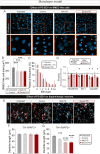
PD-EEVs reduce tight junction levels in BMEC-like cells and induce dopaminergic neuron atrophy. (A) Representative confocal images of control BMEC-like cells immunostained for VE-cadherin (white), internalized EEVs are shown in red (red, white arrows) and DAPI nuclear stain is shown in blue. Scale bar: 20 μm. (B-C) Quantification of BMEC-like cell area (B) and frequency distribution (C) in untreated conditions or after 48 h of treatment with EEV derived from control, mild PD or severe PD. (D) Western blot-based quantification of tight junction protein levels for VE-cadherin, ZO-1, and Claudin 5 in BMEC-like cell monolayers after a 48-h incubation with EEVs. Protein levels are normalized to BMEC-like cell treated with EEV control. (E) Representative confocal images of iPSC-derived neurons labeled for the pan-neuronal marker MAP2 (white) and a marker specific for dopaminergic neurons (TH, red). Images were taken after a 72-h treatment with vehicle control (i.e. untreated), age-/sex-matched control EEVs, mild or severe PD EEVs. Scale bar: 25 μm. (F-G) Quantification of MAP2+/TH (F) and TH+/MAP2+ (G) neurite lengths when iPSC-derived neurons are incubated in the absence or presence of EEVs derived from PD donors diagnosed with mild or severe PD, or age-/sex-matched controls, as shown in (E). Demographics: control subjects (n = 2–4, mean age = 70±4), patients with mild (n = 2–4, UPDRS = 26±3, mean age = 60±3) or severe (n = 4, UPDRS = 90±8, mean age = 68±5) PD. Statistical analysis: Error bars represent mean + SEM. Statistical analysis was performed using One-way ANOVA followed by Tukey’s multiple comparisons test: Comparison vs. untreated ***p-value < 0.001, ****p-value < 0.0001; Comparison vs. control EEV %%p-value < 0.01, %%%%p-value < 0.001 (B, F-G); Kuskal-Wallis test followed by Dunn’s multiple comparisons test: Comparison vs. untreated ***p-value < 0.001, ****p-value < 0.0001; Comparison vs. PD severe EEV p-value < 0.0001 (C). Statistical analysis was performed using Wilcoxon signed rank test with theoretical median (D – Ctrl EEV) set at 1 (*p-value < 0.05). (B-C) Corresponds to the BMEC-like area (B) or EEV/BMEC-like cell (C) of two independent replicates where two different iPSC donors (line IDs 38554 and 41658) and EEVs from two different individuals per group were used (i.e. at least 300 cells/experimental group); (D) Corresponds to two different iPSC donors (line IDs 38554 and 41658) and EEVs sampled from three to four different individuals per group were used. (F-G) Corresponds to the combined neurite length values of two independent replicates where two different iPSC donors (line IDs 38554 and IM2-GC) and EEVs from two different donors per group were used (i.e. at least 150 neurites/experimental group). Abbreviations: DAPI, 4′,6-diamidino-2-phenylindole; EEV, extracellular vesicles derived from erythrocytes; MAP2, microtubule-associated protein 2; ZO-1, Zonula occludens-1

Similar articles

References

    1. Spillantini MG, Schmidt ML, Lee VM, Trojanowski JQ, Jakes R, Goedert M. Alpha-synuclein in lewy bodies. Nature. 1997;388:839–40. - PubMed
    1. Lau K, Porschen LT, Richter F, Gericke B. Microvascular blood-brain barrier alterations in isolated brain capillaries of mice over-expressing alpha-synuclein (Thy1-aSyn line 61). Neurobiol Dis. 2023;187:106298. - PubMed
    1. de Rus Jacquet A, Alpaugh M, Denis HL, Tancredi JL, Boutin M, Decaestecker J, Beauparlant C, Herrmann L, Saint-Pierre M, Parent M, Droit A, Breton S, Cicchetti F. The contribution of inflammatory astrocytes to BBB impairments in a brain-chip model of Parkinson’s disease. Nat Commun. 2023;14:3651. - PMC - PubMed
    1. Al-Bachari S, Naish JH, Parker GJM, Emsley HCA, Parkes LM. Blood-Brain barrier leakage is increased in Parkinson’s disease. Front Physiol. 2020;11:593026. - PMC - PubMed
    1. Kortekaas R, Leenders KL, van Oostrom JC, Vaalburg W, Bart J, Willemsen AT, Hendrikse NH. Blood-brain barrier dysfunction in parkinsonian midbrain in vivo. Ann Neurol. 2005;57:176–9. - PubMed

MeSH terms

LinkOut - more resources