Comparative analysis of hip joint development abnormalities and risk factors in preterm and term infants
- PMID: 40229807
- PMCID: PMC11998400
- DOI: 10.1186/s40001-025-02546-y
Comparative analysis of hip joint development abnormalities and risk factors in preterm and term infants
Abstract
Objective: To investigate the risk factors affecting hip joint development in infants and to compare abnormalities in hip joint development between preterm and term infants.
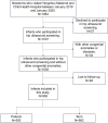
Methods: This retrospective cohort study reviewed the medical records of newborns admitted to the neonatology department of our hospital between January 2019 and January 2020. Hip joint ultrasound screening and follow-up data were collected for each newborn. The enrolled newborns were categorized into two groups: preterm (<37 weeks) and term (≥37 weeks). Hip joint ultrasounds were assessed using the Graf classification criteria.
Results: A total of 955 newborns were included in the study, comprising 393 preterm and 562 term infants. All preterm infants were born at a gestational age over 28 weeks. Among term infants, the proportion of abnormal hip joints during the first and second screenings was significantly higher in singletons than twins (p < 0.05), in non-small for gestational age (non-SGA) infants than SGA infants (p < 0.05), and in females than males (p < 0.0001). By the third screening, the proportion of abnormal hip joints remained significantly higher in females than in males (p < 0.01). In newborns with birth weight percentiles above the 50th percentile (P50), term infants showed a significantly higher proportion of abnormal hip joints than preterm infants during the first screening (P50-P75: 32.1 vs. 15.8%, P75-P90: 36.0 vs. 16.7%, p < 0.01; >P90: 32.1 vs. 15.8%, p < 0.05). During the second screening, term infants in the P50-P75 and P75-P90 percentiles exhibited abnormal hip joint proportions of 27.6 and 34.8%, respectively, which were significantly higher than those in preterm infants (13.9%, p < 0.05; 12.1%, p < 0.01). By the third screening, the proportion of abnormal hip joints in term infants within the P75-P90 percentile was 11.2%, which was significantly higher than that of preterm infants (1.5%, p < 0.05). Overall, preterm infants demonstrated a significantly lower proportion of abnormal hip joints compared to term infants across all four screening time points. By the fourth screening, all abnormal hips in preterm infants had evolved into normal ones according to Graf classification criteria. In contrast, although most abnormal hips in term infants had also resolved, with only 6 of them persisted.
Conclusion: Term singletons, female, and non-SGA infants demonstrated a higher proportion of abnormal hip joints during the early postnatal period. Among neonates born after 28 weeks of gestation, there is no difference in hip development between preterm infants with a birth weight percentile <P50 and term infants. Most abnormal hip joints in both preterm and term infants are due to physiological immaturity and tend to resolve by themselves as natural growth and development. The timing of hip ultrasound screening for preterm infants born after 28 weeks of gestation may not require correction for gestational age.
Keywords: Developmental dysplasia of the hip; Preterm infants; Risk factors; Term infants; Ultrasound screening.
© 2025. The Author(s).
Conflict of interest statement
Declarations. Ethics approval and consent to participate: This study was conducted in accordance with the relevant guidelines and the Declaration of Helsinki. It is a retrospective study of clinical data and it has been approved by the Ethics Review Committee of Yangzhou Maternal and Child Health Care Hospital Affiliated to Yangzhou University (Approval No: 202015). Because of the retrospective nature of this study, the informed consent for inclusion was waived by the ethics committee of Yangzhou Maternal and Child Health Care Hospital Affiliated to Yangzhou University. Competing interests: The authors declare no competing interests.
Figures


Similar articles
-
Breech preterm infants are at risk of developmental dysplasia of the hip.J Paediatr Child Health. 2013 Aug;49(8):658-63. doi: 10.1111/jpc.12250. Epub 2013 Jun 12. J Paediatr Child Health. 2013. PMID: 23758088
-
Does gestational age affect ultrasonographic findings of the hip in preterm newborns? A sonographic study of the early neonatal period.J Pediatr Orthop B. 2019 Mar;28(2):107-110. doi: 10.1097/BPB.0000000000000541. J Pediatr Orthop B. 2019. PMID: 30192257
-
Developmental Dysplasia of the Hip Is Not Associated with Breech Presentation in Preterm Infants.Am J Perinatol. 2024 May;41(S 01):e465-e469. doi: 10.1055/s-0042-1756139. Epub 2022 Sep 12. Am J Perinatol. 2024. PMID: 36096137
-
Cochrane Review: Screening programmes for developmental dysplasia of the hip in newborn infants.Evid Based Child Health. 2013 Jan;8(1):11-54. doi: 10.1002/ebch.1891. Evid Based Child Health. 2013. PMID: 23878122 Review.
-
Is prematurity a risk factor for developmental dysplasia of the hip? A systematic review and meta-analysis.J Pediatr Orthop B. 2023 Jul 1;32(4):305-311. doi: 10.1097/BPB.0000000000001021. Epub 2022 Nov 14. J Pediatr Orthop B. 2023. PMID: 36445370
References
-
- MacKenzie IG, Wilson JG. Problems encountered in the early diagnosis and management of congenital dislocation of the hip. J Bone Joint Surg Br. 1981;63(1):38–42. - PubMed
-
- Walton MJ, et al. The success of management with the Pavlik harness for developmental dysplasia of the hip using a United Kingdom screening programme and ultrasound-guided supervision. J Bone Joint Surg Br. 2010;92(7):1013–6. - PubMed
-
- Kishore M, et al. Detection and nonoperative management of pediatric developmental dysplasia of the hip in infants up to six months of age. J Am Acad Orthop Surg. 2015;23(3):202–5. - PubMed
Publication types
MeSH terms
Grants and funding
LinkOut - more resources
Full Text Sources
Medical
Research Materials

