. 2025 Apr 14;18(1):33.
doi: 10.1186/s13041-025-01204-y.

Development of Cre-dependent retrograde trans-multisynaptic tracer based on pseudorabies virus bartha strain

Affiliations

Development of Cre-dependent retrograde trans-multisynaptic tracer based on pseudorabies virus bartha strain

Hu You et al. Mol Brain. .

Abstract

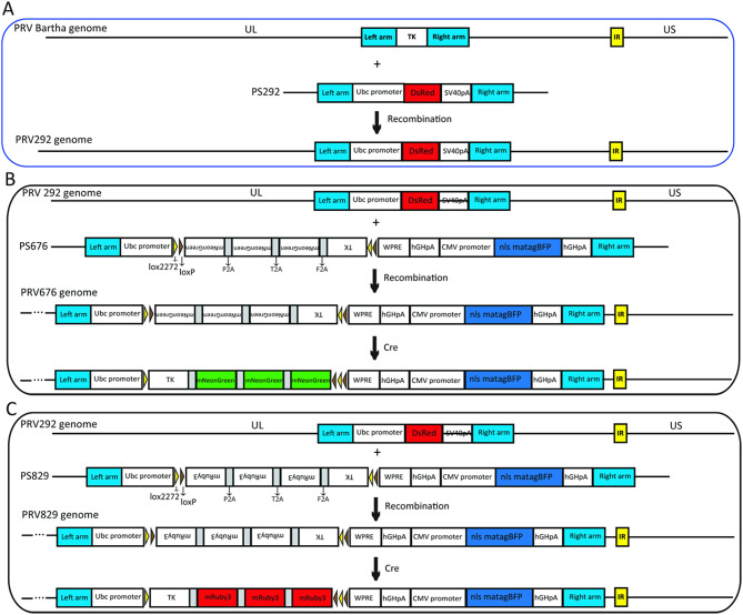
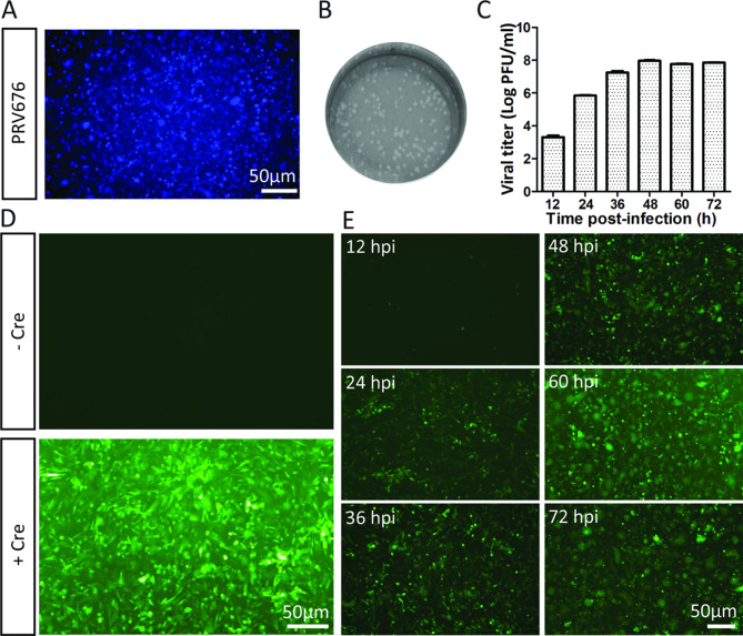
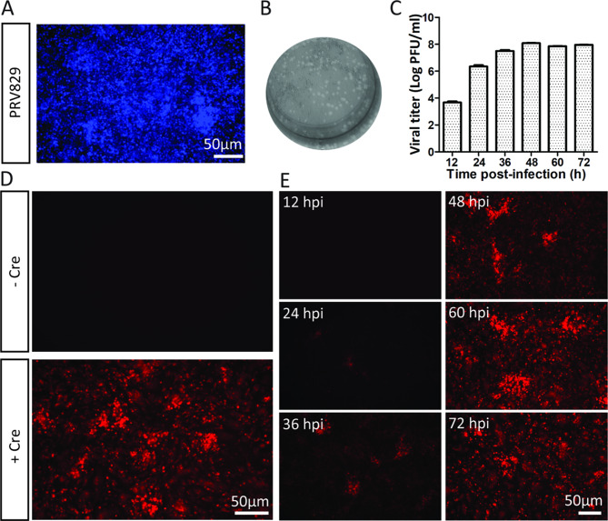
Mapping the neural circuit of a specific neuronal subclass is central to understanding the working mechanism of the brain. Currently, numerous types of transgenic mice expressing Cre recombinase have been engineered and widely used in neuroscience. To map the multilevel inputs into the neural circuit of a specific neuronal subpopulation, a Cre-dependent retrograde trans-multisynaptic tracer must be developed. The vaccine strain of Pseudorabies virus (PRV, Bartha strain) can infect neurons and spread in a retrograde manner in the neural circuit. In this study, we engineered the genome of PRV Bartha strain to prepare two new tracers, PRV676 and PRV829, by replacing the TK gene of PRV with the Cre-dependent expression cassette of the fluorescent protein gene and the TK gene. These two tracers can separately and Cre-dependently express EGFP and mRuby3 and produce progeny viruses in vitro and in vivo, which can help to map the multilevel inputs of a specific neuronal subpopulation expressing Cre. Collectively, our work provides two new tools for neuroscience research.

Keywords: Cre-dependent; PRV676; PRV829; Pseudorabies virus; Retrograde trans-multisynaptic tracer.


Conflict of interest statement

Declarations. Ethics approval and consent to participate: All procedures were approved by the Animal Care and Use Committees at the Shenzhen Institutes of Advanced Technology, Chinese Academy of Sciences. Consent for publication: Not applicable. Competing interests: The authors declare no competing interests.

Figures

Fig. 1
Fig. 1
Schematic diagram of preparation of PRV tracers. (A). The PRV292 was prepared by means of homologous recombination between PS292 and the genome of PRV Bartha strain, which was used for preparing the PRV676 and PRV829 tracers. The TK gene was deleted from the genome of PRV292. The PRV676 and PRV829 were generated by using the strategy in (B) and (C), respectively. Briefly, the PS676 and PS829 were separately tranfected into BHK-21 cells, and then the PRV292 was added into the wells. If the Cre was present in neurons, the fluoresent protein and the TK protein were expressed, and then the progeny viruses were produced
Fig. 2
Fig. 2
The characteristic of PRV676 in vitro. (A) The blue fluorescent protein (BFP) was expressed when the PRV676 infected the BHK-21 cells, which provided a direct signal for virus production in vitro. (B) The PRV676 can infect BHK-21 cells and produce plaque. (C) The growth curve of PRV676. The BHK-21 cells were infected by the PRV676 at an MOI of 0.1. The supernatant was collected at 12, 24, 36, 48, 60, and 72 hpi, and the titers of these samples were determined by using plaque assay. (D) The EGFP was expressed under the control of Cre. (E) The time course of EGFP expression and virus production in BHK-21 cells expressing Cre. The EGFP signals were observed at 12, 24, 36, 48, 60, and 72 hpi
Fig. 3
Fig. 3
The characteristic of PRV829 in vitro. (A) The blue fluorescent protein (BFP) was expressed when the PRV829 infected the BHK-21 cells, which provided a direct signal for virus production in vitro. (B) The PRV829 can infect BHK-21 cells and produce plaque. (C) The growth curve of PRV829. The BHK-21 cells were infected by the PRV829 at an MOI of 0.1. The supernatant was collected at 12, 24, 36, 48, 60, and 72 hpi, and the titers of these samples were determined by using plaque assay. (D) The mRuby3 was expressed under the control of Cre. (E) The time course of mRuby3 expression and virus production in BHK-21 cells expressing Cre. The mRuby3 signals were observed at 12, 24, 36, 48, 60, and 72 hpi
Fig. 4
Fig. 4
PRV676 and PRV829 Cre-dependently express fluorescent protein and produce progeny viruses in brain. (A) and (B) The PRV676 (9.3 × 109 PFU/ml, 300 nl) was separately injected into the VTA of the C57BL/6J mice and DAT-Cre mice. (A). The C57BL/6J mice infected with the PRV676 survived through the experimental stage, while DAT-Cre mice infected by PRV676 was dead by 6 dpi (B). (C) and (D) The PRV829 (1.8 × 109 PFU/ml, 300 nl) was separately injected into the VTA of the C57BL/6J mice and DAT-Cre mice. The mRuby3 was only observed in neurons expressing Cre (C). The C57BL/6J mice infected with the PRV829 survived through the experimental stage, while DAT-Cre mice infected by the PRV829 was dead by 6 dpi (D)
Fig. 5
Fig. 5
PRV676 can map the multilevel inputs of neurons expressing Cre in VTA of DAT-Cre mice. (A) The schematic diagram of PRV676 injection and retrograde trans-multisynaptic spreading. (B) The PRV676 (9.3 × 109 PFU/ml, 300 nl) was separately injected into the VTA of the DAT-Cre mice. The brains were treated and cut at 7 dpi. The EGFP was observed in various brain regions. BNST, bed nucleus of the stria terminalis; LPO, lateral preoptic area; MPA, medial preoptic area; VHPC, ventral hippocampus; DR, dorsal raphe nucleus; Ect, ectorhinal cortex; PRh, perirhinal cortex; MNR, median raphe nucleus; LS, lateral septal nucleus; MS, medial septal nucleus; PIR, piriform cortex; HPC, hippocampus; AMY, amygdala; LC, locus coeruleus; NDB, nucleus of the diagonal band
Fig. 6
Fig. 6
The infection of PRV676 on brain sections can not impede the molecular analysis. The PRV676 (9.3 × 109 PFU/ml, 300 nl) was injected into the VTA of the DAT-Cre mice. The brains were treated and cut at 7 dpi. The Cre (A) and NeuN (B) on brain sections were immunostained with Cre antibody and NeuN antibody, respectively. BNST, bed nucleus of the stria terminalis; LPO, lateral preoptic area; LS, lateral septal nucleus; LC, locus coeruleus; MPA, medial preoptic area
Fig. 7
Fig. 7
PRV829 can map the multilevel inputs of neurons expressing Cre in DLS of vGAT-Cre mice. (A) The schematic diagram of PRV829 injection and retrograde trans-multisynaptic spreading. (B) The PRV829 (4.5 × 109 PFU/ml, 300 nl) was separately injected into the DLS of the vGAT-Cre mice. The brains were treated and cut at 6 dpi. The mRuby3 was observed in various brain regions. HPC, hippocampus; LC, locus coeruleus; LEnt, lateral entorhinal cortex; Pa paraventricular hypothalamic nucleus; LS, lateral septal nucleus; MS, medial septal nucleus; NDB, nucleus of the diagonal band; VTA, ventral tegmental area; LPO, lateral preoptic area; MPA, medial preoptic area; VHPC, ventral hippocampus; AMY, amygdala; PIR, piriform cortex

Similar articles

References

    1. Xu X, Holmes TC, Luo MH, Beier KT, Horwitz GD, Zhao F, Zeng W, Hui M, Semler BL, Sandri-Goldin RM. Viral vectors for neural circuit mapping and recent advances in Trans-synaptic anterograde tracers. Neuron. 2020;107(6):1029–47. - PMC - PubMed
    1. Liu Q, Wu Y, Wang H, Jia F, Xu F. Viral tools for neural circuit tracing. Neurosci Bull. 2022;38(12):1508–18. - PMC - PubMed
    1. Nectow AR, Nestler EJ. Viral tools for neuroscience. Nat Rev Neurosci. 2020;21(12):669–81. - PMC - PubMed
    1. Navabpour S, Kwapis JL, Jarome TJ. A neuroscientist’s guide to Transgenic mice and other genetic tools. Neurosci Biobehav Rev. 2020;108:732–48. - PMC - PubMed
    1. Yang XW, Gong S. An overview on the generation of BAC transgenic mice for neuroscience research. Curr Protoc Neurosci 2005, Chap. 5:Unit 5 20. - PubMed

LinkOut - more resources