

Myxoid leiomyosarcoma of the uterus in a woman of childbearing age: A case report
- PMID: 40230424
- PMCID: PMC11995690
- DOI: 10.3892/ol.2025.15010
Myxoid leiomyosarcoma of the uterus in a woman of childbearing age: A case report
Abstract
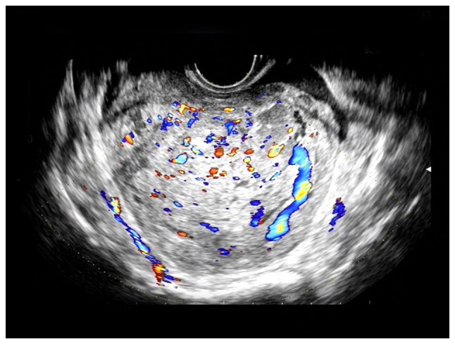
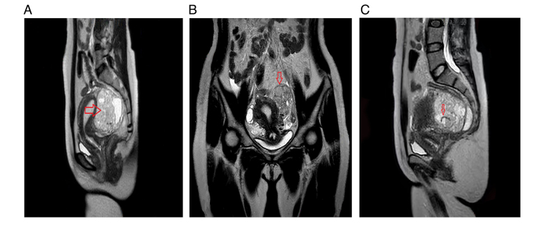
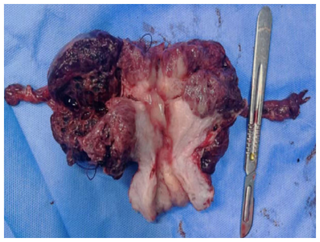
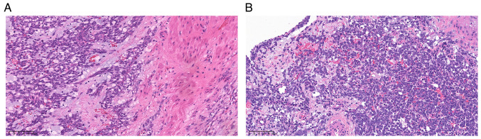
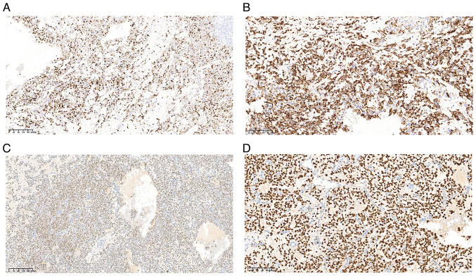
Myxoid leiomyosarcoma of the uterus (MLMS) is an extremely rare malignancy characterized by a poor prognosis. The present report describes a rare case of MLMS in a woman of childbearing age. The patient was a 33-year-old woman who presented with lower abdominal pain and was initially diagnosed with uterine myoma by preoperative B-mode ultrasound and magnetic resonance imaging. However, intraoperative pathological biopsy indicated the presence of a malignant tumor. The patient underwent total uterine and bilateral salpingectomy, along with pelvic lymph node biopsy, which confirmed the diagnosis of MLMS postoperatively. In a subsequent surgical procedure, the bilateral ovaries, omentum and appendix were also resected. The patient then received chemotherapy with gemcitabine and docetaxel and remains in good health, with no signs of recurrence. The present case report underscores the importance of early diagnosis and personalized treatment strategies for MLMS, particularly regarding their implications for fertility and survival outcomes in young women.
Keywords: case report; myxoid leiomyosarcoma; uterine myoma; women of childbearing age.
Copyright: © 2025 Wang et al.
Conflict of interest statement
The authors declare that they have no competing interests.
Figures
Similar articles
-
Uterine myxoid leiomyosarcoma with tumor embolism extending into the right atrium.Case Rep Obstet Gynecol. 2015;2015:316262. doi: 10.1155/2015/316262. Epub 2015 Feb 2. Case Rep Obstet Gynecol. 2015. PMID: 25722901 Free PMC article.
-
Uterine myxoid leiomyosarcoma initially showing low signal intensity on T2 weighted images: A case report.Radiol Case Rep. 2023 Nov 29;19(2):700-705. doi: 10.1016/j.radcr.2023.11.034. eCollection 2024 Feb. Radiol Case Rep. 2023. PMID: 38094194 Free PMC article.
-
Magnetic resonance imaging findings of a myxoid leiomyosarcoma of the uterus: A case report and literature review.Eur J Radiol Open. 2021 Feb 3;8:100328. doi: 10.1016/j.ejro.2021.100328. eCollection 2021. Eur J Radiol Open. 2021. PMID: 33604419 Free PMC article.
-
Myxoid Leiomyosarcoma of the Uterus: A Clinicopathologic Analysis of 30 Cases and Review of the Literature With Reappraisal of Its Distinction From Other Uterine Myxoid Mesenchymal Neoplasms.Am J Surg Pathol. 2016 Mar;40(3):285-301. doi: 10.1097/PAS.0000000000000593. Am J Surg Pathol. 2016. PMID: 26866354 Review.
-
Uterine myxoid leiomyosarcoma - a rare malignant tumor: the role of complex morphopathological assay. Review and case presentation.Rom J Morphol Embryol. 2021 Oct-Dec;62(4):883-896. doi: 10.47162/RJME.62.4.01. Rom J Morphol Embryol. 2021. PMID: 35673808 Free PMC article. Review.
References
-
- Chinese Anticancer Association Gynecological Tumor Professional Committee, corp-author. Diagnosis of uterine sarcoma in treatment guidelines (2021 edition) China Oncology. 2021;31:513–519. (In Chinese)
-
- Harlow BL, Weiss NS, Lofton S. The epidemiology of sarcomas of the uterus. J Natl Cancer Inst. 1986;76:399–402. - PubMed
-
- Istrate-Ofiţeru AM, Zorilă GL, Ruican D, Petrescu AM, Berbecaru EIA, Roşu GC, Căpitănescu RG, Nagy RD, Cercelaru L, Edu A, et al. Uterine myxoid leiomyosarcoma-A rare malignant tumor: The role of complex morphopathological assay. Review and case presentation. Rom J Morphol Embryol. 2021;62:883–896. doi: 10.47162/RJME.62.4.01. - DOI - PMC - PubMed
Publication types
LinkOut - more resources
Full Text Sources