. 2025 Mar 15;28(4):112120.
doi: 10.1016/j.isci.2025.112120. eCollection 2025 Apr 18.

Combination therapy with alisertib enhances the anti-tumor immunity induced by a liver cancer vaccine

Affiliations

Combination therapy with alisertib enhances the anti-tumor immunity induced by a liver cancer vaccine

Fang Xue et al. iScience. .

Abstract

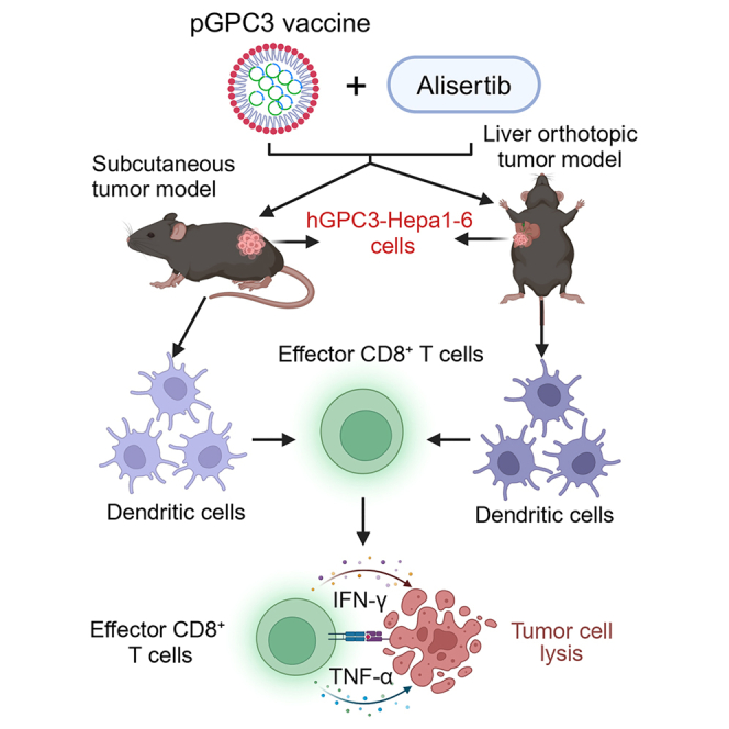
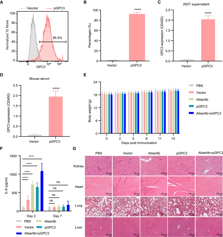
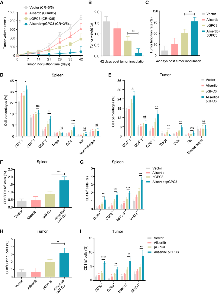
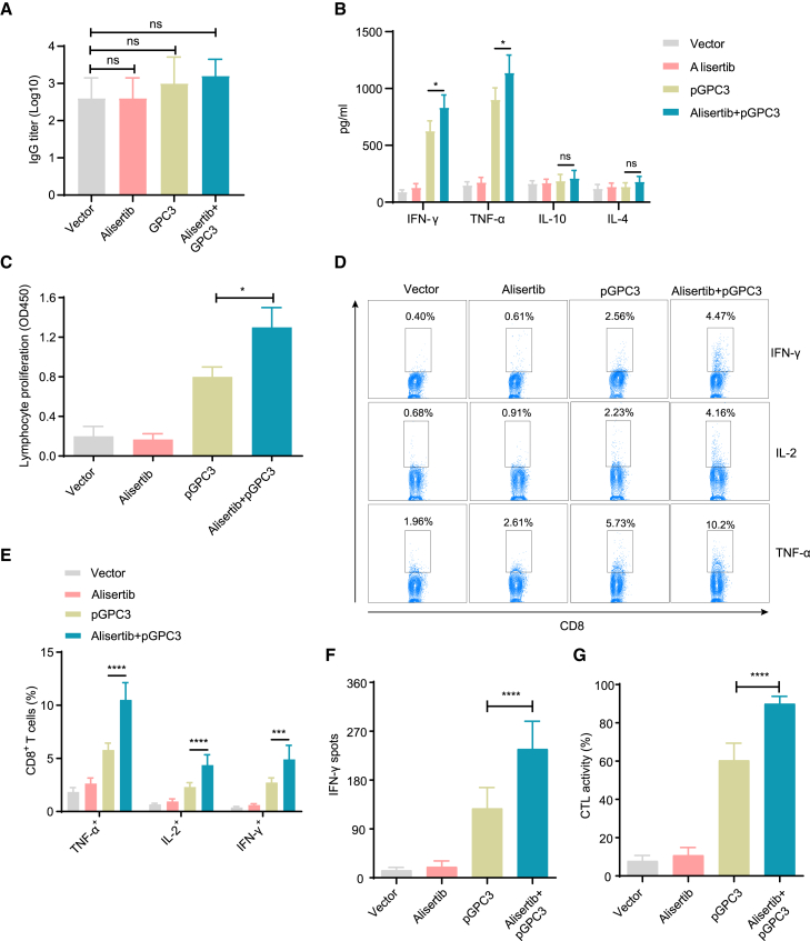
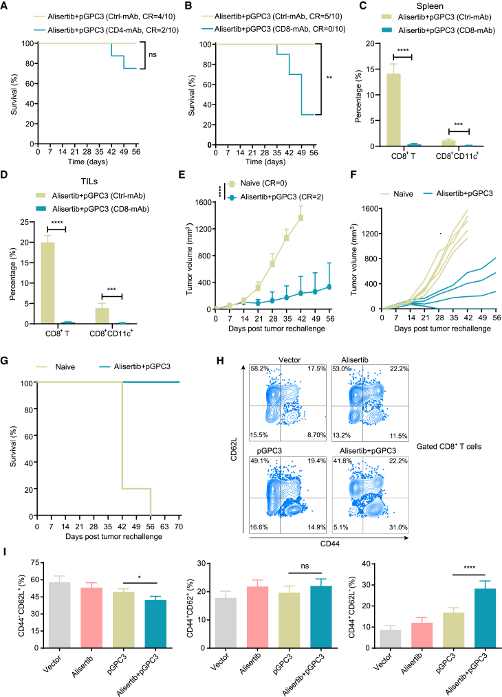
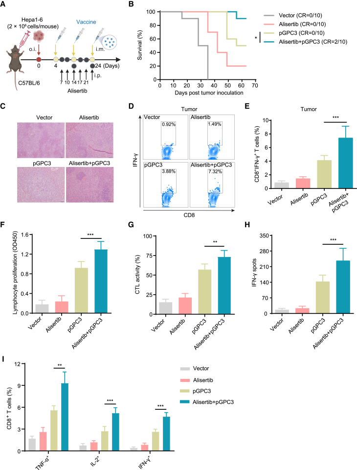
Alisertib is a potent aurora A kinase inhibitor in clinical trials for cancer treatment, but its efficacy on cancer vaccines remains unclear. Here, we developed a DNA vaccine targeting glypican-3 (pGPC3) and evaluated its efficacy with alisertib in hepatocellular carcinoma (HCC) models. The combination therapy of pGPC3 vaccine and alisertib significantly inhibited subcutaneous tumor growth, enhanced the induction and maturation of CD11c+ and CD8+CD11c+ dendritic cells (DCs), and expanded tumor-specific CD8+ T cell responses. CD8+ T cell depletion abolished the anti-tumor effects, underscoring the essential role of functional CD8+ T cell responses. Moreover, the combined treatment promoted memory CD8+ T cell induction, providing long-term protection. In liver orthotopic tumor models, the combination of pGPC3 vaccine and alisertib demonstrated potent therapeutic efficacy through CD8+ T cell responses. These results indicate that alisertib enhances the pGPC3 vaccine's therapeutic effect, offering a promising strategy for HCC treatment.

Keywords: Biological sciences; Cancer systems biology; Immunology; Natural sciences; Systems biology.


Conflict of interest statement

The authors declare no competing interests.

Figures

None
Graphical abstract
Figure 1
Figure 1
Evaluation of vaccine antigen expression and safety in the combination treatment of pGPC3 and alisertib (A) GPC3 protein expression was analyzed by flow cytometry in pGPC3-transfected 293T cells. (B) Quantification of the proportion of GPC3-positive cells from (A). (C) The protein level of GPC3 was measured by ELISA in the supernatant of pGPC3-transfected 293T cells. (D) The level of GPC3 protein was detected by ELISA in the serum of pGPC3-immunized mice. (E) Body weight changes in tumor-bearing mice were monitored every 2–3 days across the PBS, vector, alisertib, pGPC3, and alisertib + pGPC3 treatment groups. (F) Serum IL-6 levels were assessed via ELISA in mice treated with PBS, vector, alisertib, pGPC3, and the combination of alisertib and pGPC3. (G) Histopathological analysis of kidney, heart, lung and liver tissues from treated mice was performed using HE staining. Scale bar: 100 μm. All experiments were conducted with 5 mice per group. Data are presented as mean ± SD. Comparisons between two groups were conducted using a two-tailed independent Student’s t test, while multiple group comparisons were performed using ANOVA. Statistical significance was defined as ∗∗∗∗p < 0.0001; ns, not significant.
Figure 2
Figure 2
Therapy efficacy and immune cell infiltration in a subcutaneous tumor model treated with the pGPC3 vaccine and alisertib Mice were intramuscularly immunized with vector, pGPC3, on days 7, 17, and 27, or combined therapy with intraperitoneal injection of alisertib on days 10, 14, 17, 21, 24, and 27 following inoculation with hGPC3-Hepal1-6 cells. (A) Tumor volumes were measured weekly from day 7 to day 42. (B) Tumor weights were recorded on day 42 post inoculation. (C) Tumor inhibition rates were calculated for each treatment group. (D and E) Flow cytometric analysis of the percentages of CD3+ T cells, CD4+ T cells, CD8+ T cells, Tregs, DCs, NK cells, and macrophages in the spleens (n = 5) and tumors (n = 5) of mice treated with vector, alisertib, pGPC3, or the combination therapy. (F) The percentages of CD8+CD11c+ cells in the splenocytes of each group were assessed by flow cytometry. (G) Frequencies of DC subsets, including CD80+CD11c+, CD86+CD11c+, MHC-II+CD11c+, and CD40+CD11c+, in the spleens from each group. (H) The percentages of CD8+CD11c+ cells in the tumors from each group were detected by flow cytometry. (I) Frequencies of CD80+CD11c+, CD86+CD11c+, MHC-II+CD11c+, and CD40+CD11c+ DC subsets in tumors across all groups. All experiments were performed with 5 mice per group. Data are presented as mean ± SD. Multiple group comparisons were performed using ANOVA. Statistical significance was set at ∗p < 0.05, ∗∗p < 0.01, ∗∗∗p < 0.001, and ∗∗∗∗p < 0.0001; ns, not significant.
Figure 3
Figure 3
Humoral and cellular immune responses induced by pGPC3 and alisertib in a subcutaneous tumor model (A) Anti-hGPC3 total IgG antibody levels in serum were measured by ELISA at day 35 post tumor inoculation. (B) The expression levels of IFN-γ, TNF-α, IL-10, and IL-4 were quantified in the culture supernatants of splenocytes stimulated with hGPC3 protein (10 μg/mL) for 48 h using ELISA. (C) Splenocytes isolated from treated mice (n = 5) were stimulated in vitro with IL-2 (100 U/mL) and hGPC3 protein (10 μg/mL) for 72 h; the cell proliferation assay was conducted and presented as optical density (OD) values. (D) Splenocytes in RPMI 1640 complete medium containing IL-2 (100 U/mL) were stimulated with hGPC3 protein (10 μg/mL) for 72 h, followed by ionomycin (500 ng/mL), PMA (50 ng/mL), and BFA (5 μg/mL) for the final 5 h. Flow cytometry was used to analyze TNF-α, IL-2, and IFN-γ-expressing CD8+ T cells in each group. (E) Statistical analysis of TNF-α, IL-2, and IFN-γ-expressing CD8+ T cell percentages from (D). (F) The number of IFN-γ-secreting CD8+ T cells was measured using the ELISPOT assay. (G) CTL activity was assessed using a co-culture-killing assay. All experiments were performed with 5 mice per group. Data are presented as mean ± SD. Multiple group comparisons were performed using ANOVA. Statistical significance was set at ∗p < 0.05, ∗∗∗p < 0.001, and ∗∗∗∗p < 0.0001; ns, not significant.
Figure 4
Figure 4
Memory CD8+ T cells are essential for the long-term anti-tumor effect of pGPC3 and alisertib (A and B) Survival curves of tumor-bearing mice treated with pGPC3 and alisertib after depletion of CD4+ T or CD8+ T cells. (C and D) Percentages of CD8+ T cells and CD8+CD11C+ subsets in splenocytes and TILs, measured by flow cytometry. (E) Tumor growth in cured mice after rechallenge with tumor cells. (F) Individual tumor growth curves for each mouse in different groups were monitored up to 56 days post rechallenge; naive mice served as controls. (G) Survival curves comparing rechallenged mice to naive controls (n = 5). (H) Flow cytometry dot plots showing CD8+ memory T cell populations in splenocytes from four experimental groups (n = 5). (I) Statistical analysis of the proportions of effector memory (CD62LCD44+), central memory (CD44+CD62L+), and naive (CD44CD62L+) CD8+ T cells in splenocytes from treated mice. All experiments were performed with 5 mice per group. Data are presented as mean ± SD. Comparisons between two groups were performed using a two-tailed independent Student’s t test. Multiple group comparisons were performed using ANOVA. Survival analysis was conducted using the log rank (Mantel-Cox) test. Statistical significance was set at ∗p < 0.05, ∗∗p < 0.01, ∗∗∗p < 0.001, and ∗∗∗∗p < 0.0001; ns, not significant.
Figure 5
Figure 5
The combination therapy of pGPC3 and alisertib inhibited liver tumor progression (A) Mice were orthotopically inoculated with Hepa1-6 cells on day 0 and treated with vector or pGPC3 on days 4, 14, and 24, alisertib or alisertib + pGPC3 on days 7, 10, 14, 17, 21, and 24 post tumor inoculation. (B) Survival of liver tumor-bearing mice was monitored up to 63 days after inoculation. (C) HE staining of liver sections showing tumor foci from representative mice in each treatment group. (D and E) Flow cytometric analysis of the percentages of tumor-infiltrating CD8+ T cells in each group. (F) Lymphocyte proliferation. (G) CTL activity analysis. (H) Quantification of IFN-γ-secreting T cells using ELISPOT assay. (I) Flow cytometric analysis of the percentages of TNF-α, IL-2, and IFN-γ-producing CD8+ T cells in stimulated splenocytes from each treatment group. Survival experiments involved 10 mice per group, while other assays were conducted with 5 mice per group. Data are presented as mean ± SD. Survival analysis was performed using the log rank (Mantel-Cox) test. Multiple group comparisons were performed using ANOVA. Statistical significance was set at ∗p < 0.05, ∗∗p < 0.01, and ∗∗∗p < 0.001.

Similar articles

References

    1. Lin M.J., Svensson-Arvelund J., Lubitz G.S., Marabelle A., Melero I., Brown B.D., Brody J.D. Cancer vaccines: the next immunotherapy frontier. Nat. Cancer. 2022;3:911–926. doi: 10.1038/s43018-022-00418-6. - DOI - PubMed
    1. Saxena M., van der Burg S.H., Melief C.J.M., Bhardwaj N. Therapeutic cancer vaccines. Nat. Rev. Cancer. 2021;21:360–378. doi: 10.1038/s41568-021-00346-0. - DOI - PubMed
    1. Baharom F., Ramirez-Valdez R.A., Khalilnezhad A., Khalilnezhad S., Dillon M., Hermans D., Fussell S., Tobin K.K.S., Dutertre C.A., Lynn G.M., et al. Systemic vaccination induces CD8(+) T cells and remodels the tumor microenvironment. Cell. 2022;185:4317–4332.e15. doi: 10.1016/j.cell.2022.10.006. - DOI - PMC - PubMed
    1. Lopes A., Bastiancich C., Bausart M., Ligot S., Lambricht L., Vanvarenberg K., Ucakar B., Gallez B., Préat V., Vandermeulen G. New generation of DNA-based immunotherapy induces a potent immune response and increases the survival in different tumor models. J. Immunother. Cancer. 2021;9 doi: 10.1136/jitc-2020-001243. - DOI - PMC - PubMed
    1. van der Burg S.H., Arens R., Ossendorp F., van Hall T., Melief C.J.M. Vaccines for established cancer: overcoming the challenges posed by immune evasion. Nat. Rev. Cancer. 2016;16:219–233. doi: 10.1038/nrc.2016.16. - DOI - PubMed

LinkOut - more resources