. 2024 Sep 8:23:15353508241270405.
doi: 10.1177/15353508241270405. eCollection 2024 Jan-Dec.

Preliminary Exploration of Al18F-NOTA-FAPI-04 PET/CT in the Management of Ankylosing Spondylitis: A Prospective Clinical Study

Affiliations

Preliminary Exploration of Al18F-NOTA-FAPI-04 PET/CT in the Management of Ankylosing Spondylitis: A Prospective Clinical Study

Shibo Guo et al. Mol Imaging. .

Abstract

Background: Ankylosing spondylitis (AS) is characterized by inflammation and osteoblastic changes in the sacroiliac joint. As a potential imaging method for the early assessment of AS, positron emission tomography (PET) can quantify systemic disease activity, which is conducive to monitoring the progression of disease activity and assisting in evaluating the efficacy of the treatment.

Objective: The study was to evaluate the diagnostic value of aluminium-[18F]fuoride(Al18F)-labelled fibroblast activation protein inhibitor (FAPI) PET/computed tomography (CT) in AS and to investigate its ability to assess disease activity during the development of AS.

Material and methods: Twenty AS participants who met the Assessment of SpondyloArthritis international Society criteria and were in an active disease stage were included in this study from May 2022 to April 2023. Sixteen healthy controls were also inrolled. All participants underwent Al18F-NOTA-FAPI-04 PET/CT imaging after collecting clinical assessment and laboratory results. The correlation between positive joint count (PJC) and systemic joint standard uptake value ratio (SUVR, the mean SUVmax of the 5 highest joints/SUVmax of the uninvolved sacrum) on PET and clinical disease activity assessment and various laboratory tests were analyzed.

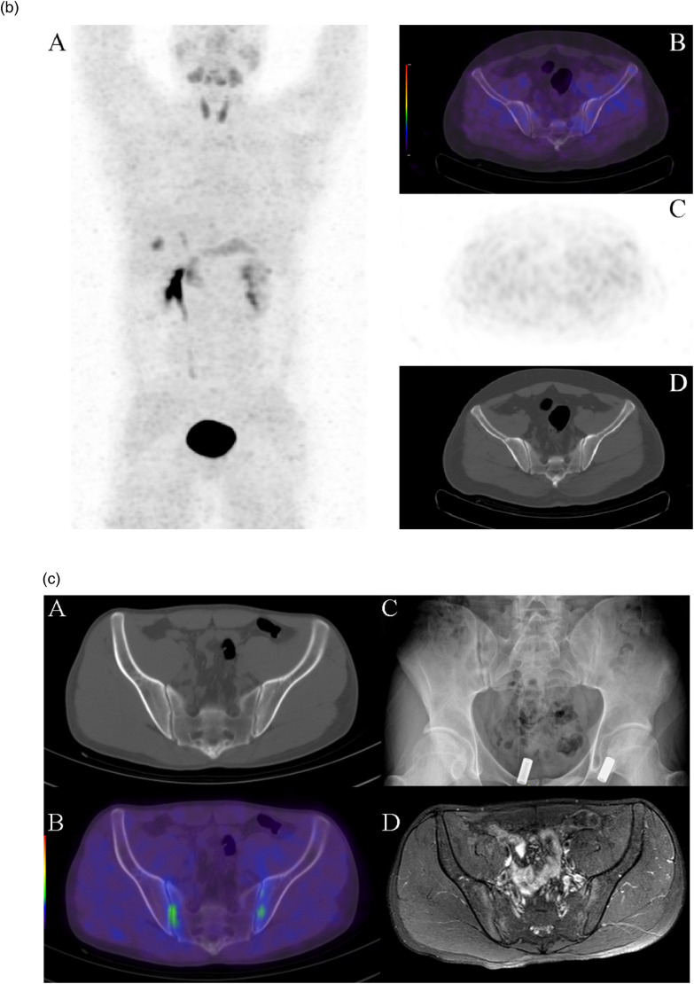
Results: A total of 2820 joints were observed in 20 participants (median age 34.5,[21-61]range, 15 men), with a PJC of 1300 (46.7%), and 39 positive uptakes were found in 40 sacroiliac joints (97.5%). PET/CT images revealed FAPI-04 uptake in both sacroiliac joints in 2 participants without radiographic sacroiliitis in the early stages of AS and varying degrees of uptake in the sacroiliac joints and spinal joints in the remaining participants. PJC and SUVR were positively correlated with most clinical assessments and laboratory findings (P < .05), and SUVR of the sacroiliac joint was positively correlated with C-reactive protein (CRP) (mg/L; r = 0.498, P = .026).

Conclusion: Al18F-NOTA-FAPI-04 PET/CT was highly sensitive to systemic arthritic changes in AS participants and correlated with clinical disease activity and laboratory tests.

Keywords: PET/CT; ankylosing spondylitis; fibroblast activation protein; inflammation.


Conflict of interest statement

The authors declared no potential conflicts of interest with respect to the research, authorship, and/or publication of this article.

Figures

Figure 1.
Figure 1.
Flow chart of the patient recruitment criteria and study procedure. Abbreviations: Al18F-NOTA-FAPI-04, aluminum-[18F]-labeled 14,7-triazacyclononaneN,N′,N″-triacetic acid-conjugated fibroblast activation protein inhibitor 04; AS, ankylosing spondylitis; ASDAS, Ankylosing Spondylitis Disease Activity Score; BASDAI, The Bath Ankylosing Spondylitis Disease Activity Index; CRP, C-reactive protein; ESR, erythrocyte sedimentation rate; HLA-B27, human leukocyte antigen B27; SAPHO, synovitis, acne, pustulosis, hyperostosis, and osteitis.
Figure 2.
Figure 2.
(a) A 28-year-old male patient with sacroiliac and low back pain of gradual worsening for 1 year, HLA-B27 positive, C-reactive protein 32.68, erythrocyte sedimentation rate 17, SUVR 3.51.ASDAS-CRP 4.2, whole-body maximum projection MIP image (A) shows bilateral sacroiliac joint and multiple vertebral joint uptake (arrows), and cross-sectional images (B-D) show bilateral sacroiliac joint radiotracer uptake with a SUVmax of 6.03, suggesting bilateral sacroiliac arthritis. (b) A 27-year-old healthy patient with Al18F-NOTA-FAPI-04 positron emission tomography images and a negative serological human leukocyte antigen B27 test did not meet the Ankylosing Spondylitis International Association criteria for Assessing Spondylitis. Maximum intensity projection image (A) and transverse axial image of the sacroiliac joint (B-D) are indicated as normal scans. (c) A 21-year-old male patient with malaise and sacroiliac joint pain for more than 1 month, HLA-B27 positive, CRP 37, erythrocyte sedimentation rate 39, SUVR 2.41, ASDAS-CRP 3.26. CT and radiographs showed no abnormalities in the sacroiliac joints with a radiological grade of 0 (A and C), and PET showed radiological aggregates in the sacroiliac joints bilaterally with a SUVmax of 2.79 (B), suggestive of bilateral sacroiliac arthritis, which was consistent with that shown by MRI (D). Abbreviations: Al18F-NOTA-FAPI-04, aluminum-[18F]-labeled 14,7-triazacyclononaneN,N′,N″-triacetic acid-conjugated fibroblast activation protein inhibitor 04; ASDAS, Ankylosing Spondylitis Disease Activity Score; CRP, C-reactive protein; CT, computed tomography; HLA-B27, human leukocyte antigen B27; MRI, magnetic resonance imaging; PET, positron emission tomography; SUVR, standard uptake value ratio.
Figure 2.
Figure 2.
(a) A 28-year-old male patient with sacroiliac and low back pain of gradual worsening for 1 year, HLA-B27 positive, C-reactive protein 32.68, erythrocyte sedimentation rate 17, SUVR 3.51.ASDAS-CRP 4.2, whole-body maximum projection MIP image (A) shows bilateral sacroiliac joint and multiple vertebral joint uptake (arrows), and cross-sectional images (B-D) show bilateral sacroiliac joint radiotracer uptake with a SUVmax of 6.03, suggesting bilateral sacroiliac arthritis. (b) A 27-year-old healthy patient with Al18F-NOTA-FAPI-04 positron emission tomography images and a negative serological human leukocyte antigen B27 test did not meet the Ankylosing Spondylitis International Association criteria for Assessing Spondylitis. Maximum intensity projection image (A) and transverse axial image of the sacroiliac joint (B-D) are indicated as normal scans. (c) A 21-year-old male patient with malaise and sacroiliac joint pain for more than 1 month, HLA-B27 positive, CRP 37, erythrocyte sedimentation rate 39, SUVR 2.41, ASDAS-CRP 3.26. CT and radiographs showed no abnormalities in the sacroiliac joints with a radiological grade of 0 (A and C), and PET showed radiological aggregates in the sacroiliac joints bilaterally with a SUVmax of 2.79 (B), suggestive of bilateral sacroiliac arthritis, which was consistent with that shown by MRI (D). Abbreviations: Al18F-NOTA-FAPI-04, aluminum-[18F]-labeled 14,7-triazacyclononaneN,N′,N″-triacetic acid-conjugated fibroblast activation protein inhibitor 04; ASDAS, Ankylosing Spondylitis Disease Activity Score; CRP, C-reactive protein; CT, computed tomography; HLA-B27, human leukocyte antigen B27; MRI, magnetic resonance imaging; PET, positron emission tomography; SUVR, standard uptake value ratio.
Figure 3.
Figure 3.
CT and PET/CT images of 5 patients with different radiological grades of sacroiliac joints. Abbreviations: CT, computed tomography; PET, positron emission tomography.
Figure 4.
Figure 4.
Bar chart comparing differences between groups of PJC and SUVR between different levels of disease activity. Two sets of bar graphs show clear trends, using one-way ANOVA, P < .05 was statistically significant. Abbreviations: ANOVA, analysis of variance; PJC, positive joint count; SUVR, standard uptake value ratio.
Figure 5.
Figure 5.
Baseline and post-treatment Al18F-NOTA-FAPI-04 PET/CT images of 3 patients, patient #3 is a 35-year-old male with an initial diagnosis of ankylosing spondylitis who was treated with adalimumab and reviewed 7 months later, patient #8 is a 36-year-old man who had been diagnosed with ankylosing spondylitis 1 month earlier and was followed up 6 months after treatment with adalimumab. Patient #10 is a 28-year-old woman with an initial diagnosis of ankylosing spondylitis. She was treated with adalimumab and followed up 6 months later. Abbreviations: Al18F-NOTA-FAPI-04, aluminum-[18F]-labeled 14,7-triazacyclononaneN,N′,N″-triacetic acid-conjugated fibroblast activation protein inhibitor 04; CT, computed tomography; PET, positron emission tomography.

Similar articles

References

    1. Sieper J, Poddubnyy D. Axial spondyloarthritis. The Lancet. 2017;390(10089):73-84. doi:10.1016/s0140-6736(16)31591-4 - DOI - PubMed
    1. Qu F, Guilak F, Mauck RL. Cell migration: implications for repair and regeneration in joint disease. Nat Rev Rheumatol. 2019;15(3):167-179. doi:10.1038/s41584-018-0151-0 - DOI - PMC - PubMed
    1. Maksymowych WP, Morency N, Conner-Spady B, Lambert RG. Suppression of inflammation and effects on new bone formation in ankylosing spondylitis: evidence for a window of opportunity in disease modification. Ann Rheum Dis. 2013;72(1):23-28. doi:10.1136/annrheumdis-2011-200859 - DOI - PubMed
    1. Braun J, Baraliakos X, Hermann KG, et al. The effect of two golimumab doses on radiographic progression in ankylosing spondylitis: results through 4 years of the GO-RAISE trial. Ann Rheum Dis. 2014;73(6):1107-1113. doi:10.1136/annrheumdis-2012-203075 - DOI - PMC - PubMed
    1. Rudwaleit M, van der Heijde D, Landewe R, et al. The development of assessment of SpondyloArthritis International Society classification criteria for axial spondyloarthritis (part II): validation and final selection. Ann Rheum Dis. 2009;68(6):777-783. doi:10.1136/ard.2009.108233 - DOI - PubMed

Substances

LinkOut - more resources