

Role of Artificial Intelligence in Congenital Heart Disease and Interventions
- PMID: 40230672
- PMCID: PMC11993855
- DOI: 10.1016/j.jscai.2025.102567
Role of Artificial Intelligence in Congenital Heart Disease and Interventions
Abstract
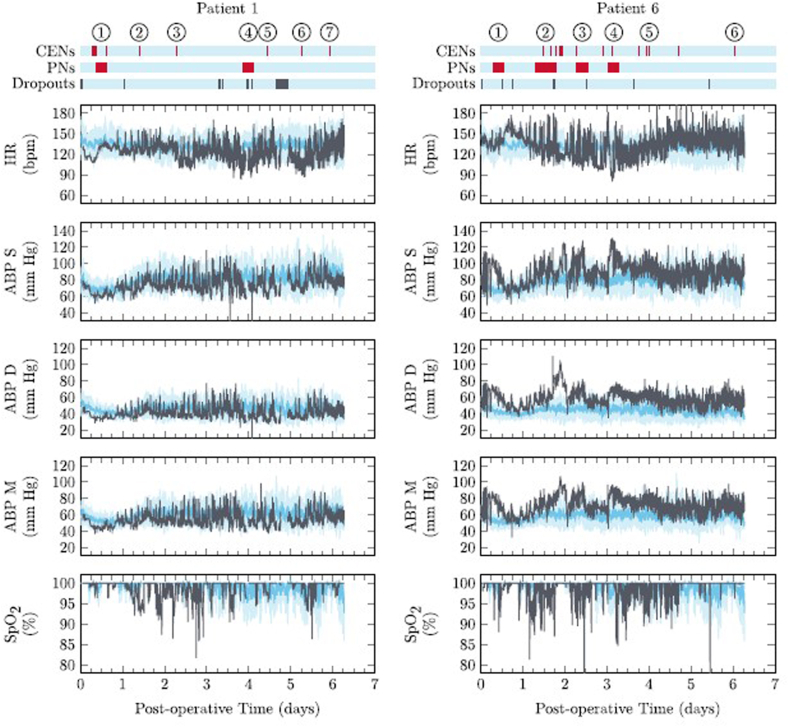
Artificial intelligence has promising impact on patients with congenital heart disease, a vulnerable population with life-long health care needs and, often, a substantially higher risk of death than the general population. This review explores the role artificial intelligence has had on cardiac imaging, electrophysiology, interventional procedures, and intensive care monitoring as it relates to children and adults with congenital heart disease. Machine learning and deep learning algorithms have enhanced not only imaging segmentation and processing but also diagnostic accuracy namely reducing interobserver variability. This has a meaningful impact in complex congenital heart disease improving anatomic diagnosis, assessment of cardiac function, and predicting long-term outcomes. Image processing has benefited procedural planning for interventional cardiology, allowing for a higher quality and density of information to be extracted from the same imaging modalities. In electrophysiology, deep learning models have enhanced the diagnostic potential of electrocardiograms, detecting subtle yet meaningful variation in signals that enable early diagnosis of cardiac dysfunction, risk stratification of mortality, and more accurate diagnosis and prediction of arrhythmias. In the congenital heart disease population, this has the potential for meaningful prolongation of life. Postoperative care in the cardiac intensive care unit is a data-rich environment that is often overwhelming. Detection of subtle data trends in this environment for early detection of morbidity is a ripe avenue for artificial intelligence algorithms to be used. Examples like early detection of catheter-induced thrombosis have already been published. Despite their great promise, artificial intelligence algorithms are still limited by hurdles such as data standardization, algorithm validation, drift, and explainability.
Keywords: artificial intelligence; congenital heart disease; deep learning; interventional cardiology; machine learning; structural heart disease.
© 2025 The Author(s).
Figures


References
Publication types
LinkOut - more resources
Full Text Sources
