. 2025 Mar 31:16:1565797.
doi: 10.3389/fimmu.2025.1565797. eCollection 2025.

CD121b-positive neutrophils predict immunosuppression in septic shock

Affiliations

CD121b-positive neutrophils predict immunosuppression in septic shock

Jian Chen et al. Front Immunol. .

Abstract

Background: Septic shock is linked with high mortality and significant long-term morbidity in survivors. However, the specific role of neutrophils in septic shock pathophysiology remains scarce in recent research.

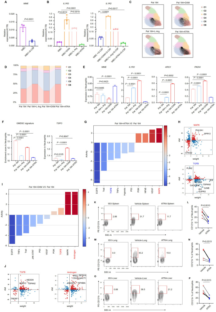
Methods: Peripheral blood immune cells from healthy donors and patients with septic shock were analyzed using single-cell RNA sequencing and batch RNA sequencing. We measured serum CD121b in both patients and healthy donors. Peripheral immune cells were isolated and exposed to either a CD121b recombinant protein or a CD121b blocking antibody to evaluate the expression of inflammatory factors. Additionally, in a humanized mouse sepsis model, the expression of CD121b in neutrophils across different tissues was assessed following treatment with all-trans retinoic acid (ATRA).

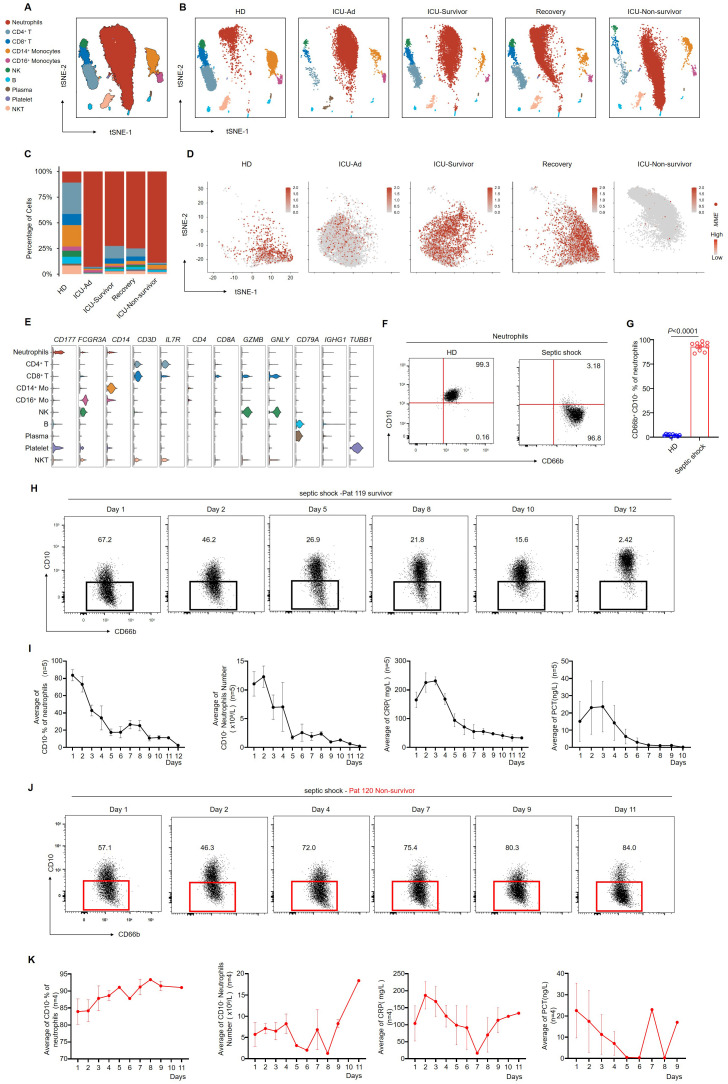
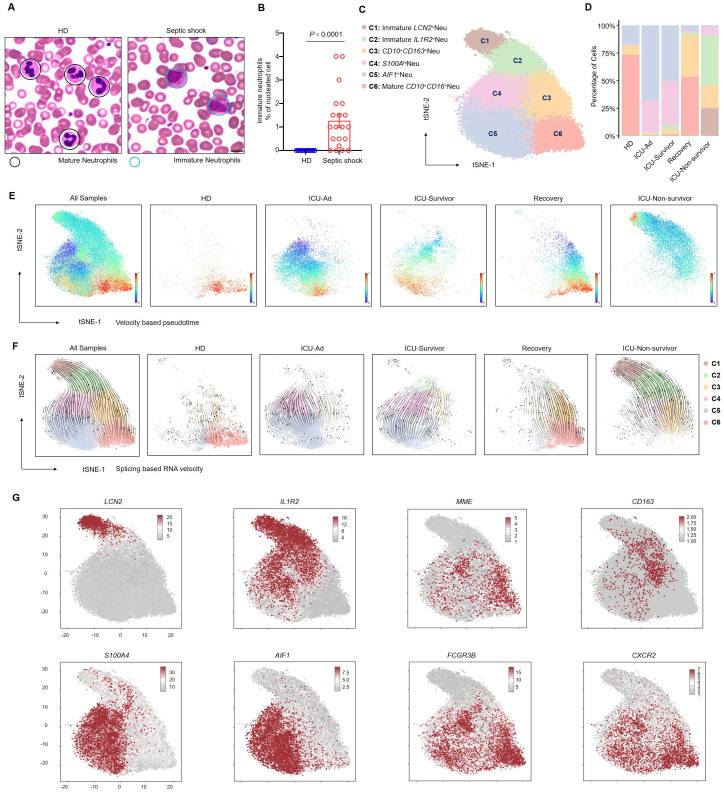
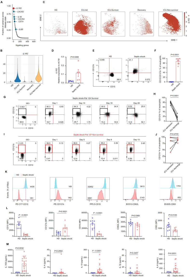
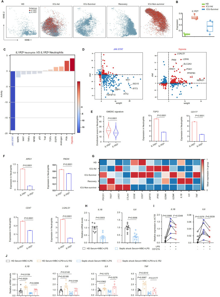
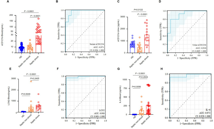
Results: This study identified a subset of CD10-CD121b+ neutrophils in the peripheral blood of patients with septic shock. These patients exhibited elevated concentrations of soluble CD121b in serum and urine. Furthermore, outcomes revealed that the presence of CD121b+ neutrophils positively correlated with the severity of septic shock. These cells displayed immunosuppressive characteristics; after blocking CD121b, proinflammatory cytokines increased in peripheral immune cells. Additionally, we found that treatment with ATRA down-regulated the expression of CD121b.

Conclusions: CD121b is closely associated with the progression of septic shock and may serve as a potential predictor indicator of immunosuppression for the condition.

Keywords: CD121b; cytokine; immunosuppression; neutrophils; septic shock.


Conflict of interest statement

The authors declare that the research was conducted in the absence of any commercial or financial relationships that could be construed as a potential conflict of interest.

Figures

Figure 1
Figure 1
CD66b+ CD10 neutrophils are related to the progression of septic shock. (A) the t-SNE plot of single-cell sequencing data (33,472 cells) from peripheral blood of patients with septic shock and HDs, annotated by distinct cell populations. (B) t-SNE plots for individual sample groups. HDs, n = 3; ICU Admission (ICU-Ad) & ICU-Survivor, n = 1; Recovery, n = 1; ICU Non-survivor, n = 1. (C) Changes in the proportions of immune cell subpopulations across conditions. (D) MME expression pattern projected on the t-SNE plot, showing neutrophils only. (E) Violin plots displaying a scaled expression of selected signature genes across clusters. (F) Representative flow cytometry plots showing the frequencies of CD66b+CD10 neutrophils in healthy donors (left) and a patient with septic shock (right). (G) Quantification of CD66b+CD10 neutrophils in healthy donors (n = 10) and septic shock patients (n = 10). (H) Dot plots showing dynamic changes in the proportion of CD66b+CD10- neutrophils in surviving septic shock patients. (I) Dynamic changes in the average proportion of CD10 neutrophils, absolute counts of CD10 neutrophils, serum CRP, and PCT levels in septic shock survivors (n = 5). (J) Dot plots showing dynamic changes in the proportion of CD66b+CD10- neutrophils in septic shock patients who died. (K) Dynamic changes in the average proportion of CD10- neutrophils among total neutrophils, absolute CD10 neutrophil counts, serum CRP, and PCT levels in patients who succumbed to septic shock (n = 4). Three or more independent experiments were performed. Statistical analyses were performed using two-tailed unpaired Student’s t-test (G).
Figure 2
Figure 2
Blockade of the development of CD66b+CD10+ neutrophils in septic shock. (A) Representative Wright–Giemsa–stained peripheral blood smears showing mature neutrophils in a healthy donor (left) and immature neutrophils in a patient with septic shock (right); scale bar, 10 μm. (B) The ratio of the proportion of immature neutrophils to nucleated cells in the peripheral blood of healthy donors (n = 10) and septic shock patients (n = 20). (C) t-SNE plot identifying six neutrophil clusters. (D) Proportions of neutrophil clusters across five patient groups. (E, F) Latent time analysis (E) and neutrophil RNA velocity (F) are displayed for each sample group on the t-SNE plot. (G) t-SNE plots illustrating the expression of selected marker genes across six neutrophil clusters. Statistical analyses were performed using the Mann–Whitney test (B).
Figure 3
Figure 3
CD10 CD121b+ neutrophils are positively correlated with mortality risk in septic shock. (A) Differential gene expression in neutrophils across distinct phases of sepsis, organized by the magnitude of change, with IL1R2 showing the most pronounced alteration. (B) Violin plot depicting IL1R2 expression in neutrophils across different phases of sepsis and healthy donors. (C) IL1R2 expression projected on the t-SNE plot, highlighting neutrophils. (D) IL1R2 expression in peripheral blood neutrophils from HDs (n = 5) and patients with septic shock (n = 5). (E) Representative flow cytometry plots showing CD10CD121b+neutrophil frequencies in a healthy donor (left) and a patient with septic shock (right). (F) Quantification of CD10 CD121b+ neutrophils in healthy donors (n = 7) and septic shock patients (n = 11). (G, H) Representative flow cytometry plots (G) and statistical analysis (H) showing the dynamics of CD10 CD121b+ neutrophil frequencies in septic shock survivors from ICU admission to discharge (n = 6). (I, J) Representative flow cytometry plots (I) and statistical analysis (J) of CD10 CD121b+ neutrophils in patients with septic shock leading to death (n = 6). (K) Histograms of CD10, CD121b, CD16, CD62L, and CD63 expression in peripheral blood neutrophils from healthy donors (upper) and septic shock patients (lower). (L) Mean fluorescence intensity (MFI) of CD10, CD121b, CD16, CD62L, and CD63 in neutrophils from healthy donors (n = 8) and septic shock patients (n = 8). (M) Serum concentrations of IL-RA, IL-4, IL-10, IL-1β, and IL-8 in healthy donors (n = 6) and septic shock patients (n = 20). Three or more independent experiments were conducted. Statistical analyses were performed using two-tailed unpaired Student’s t-test (D, F, K), paired t-test (H, J), and Mann–Whitney test (L).
Figure 4
Figure 4
CD121bhi neutrophils have an immunosuppression role in septic shock. (A) t-SNE plot displaying two distinct neutrophil populations separated by IL1R2 expression across five sample groups. (B) Based on RNA-sequencing data, a bar graph illustrating gene-signature enrichment scores of highly expressed IL1R2 in neutrophils. (C) Inferred pathway activity among neutrophil subclusters using the PROGENy platform. (D) Differentially expressed genes specific to the JAK-STAT and hypoxia signaling pathways. (E) Enrichment scores of the GMDSC signature in neutrophil subclusters, with bar graphs showing variation in the expression of TSPO and CD177. (F) Bar graphs depicting variation in the expression of immunosuppressive genes in neutrophils. (G) Heatmap displaying a differential expression of immune-related genes in T cells across five sample groups. (H) Gene expression of IL1B and IL6 in white blood cells from HDs following 24 h of LPS stimulation in serum from HDs (n = 8) or septic shock patients (n = 8). (I) Gene expression of IL1B and IL6 in white blood cells from HDs (n = 6) following 24 h of LPS stimulation in FBS with or without recombinant human CD121b protein. (J) Gene expression of IL1B, IL6, IL8, and TNF in white blood cells from HDs following 24 h of LPS stimulation in serum from HDs (n = 5) or septic shock patients (n = 5) with or without the addition of CD121b (IL-1R2)-blocking antibodies. Three or more independent experiments were performed. Statistical analyses were conducted using two-tailed unpaired Student’s t-test (E, F, H), paired t-test (I, J), and Mann-Whitney test (J).
Figure 5
Figure 5
ATRA alleviates the immunosuppressive effect of neutrophils. (A) MME expression in white blood cells from septic shock patients (n = 3) after 24 h of G-CSF stimulation. (B) IL1R2 expression in white blood cells from septic shock patients (n = 3) following 24 h of stimulation with dexamethasone, L-arginine, or ATRA, with or without G-CSF. (C) Single-cell sequencing of neutrophils from septic shock patients after in vitro stimulation with DXM, L-Arg, or ATRA, with neutrophil subpopulations displaying maturation features circled in black. (D) Changes in the proportions of neutrophil subclusters. (E) Bar graphs showing variation in the expression of key genes in neutrophils of single-cell sequencing data. (F) GMDSC signature enrichment in neutrophils, with a bar graph showing variation in TSPO expression of single-cell sequencing data. (G, H) Pathway activity changes (G) and differentially expressed genes (H) in the MAPK and TGF-β signaling pathways in neutrophils treated with ATRA. (I, J) Pathway activity changes (I) and differentially expressed genes (J) in the TGF-β and androgen signaling pathways in neutrophils treated with DXM. (K-O) Representative flow cytometry plots showing the proportion of CD121b+ human neutrophils in the spleen (K), lung (M), and liver (O) of NCG mice 24 h after ATRA administration. (L, N, P) Statistical analysis of CD121b+ human neutrophil proportions in the spleen (L), lung (N), and liver (P) of vehicle-treated (n = 6) and ATRA-treated (n = 6) NCG mice. Three or more independent experiments were performed. Statistical analyses were conducted using two-tailed unpaired Student’s t-test (A, E, F), one-way ANOVA (B), paired t-test (L), and Wilcoxon test (N, P).
Figure 6
Figure 6
CD121b as a biomarker for the immunosuppressive state in patients with septic shock. (A) Serum levels of sCD121b in HDs (n = 57), non-septic patients (n = 14), and septic shock patients (n = 39). (B) The ROC curve for serum CD121b levels comparing non-septic patients and septic shock patients. (C) Urine levels of sCD121b in HDs (n = 20), non-septic patients (n = 14), and septic shock patients (n = 15). (D) ROC curve for urine CD121b levels comparing non-septic and septic shock patients. (E) Serum levels of LCN2 in HDs (n = 10), non-septic patients (n = 13), and septic shock patients (n = 13). (F) ROC curve for serum LCN2 levels comparing HDs and sepsis patients (including non-septic and septic shock patients). (G) Serum levels of IL-6 in HDs (n = 10), non-septic patients (n = 14), and septic shock patients (n = 39). (H) ROC curve for serum IL-6 levels comparing HDs and sepsis patients (including non-septic and septic shock patients). Three or more independent experiments were performed. Statistical analyses were performed using the Kruskal-Wallis test (A, C, E, G) and AUC analysis (B, D, F, H).

Similar articles

References

    1. Hotchkiss RS, Monneret G, Payen D. Sepsis-induced immunosuppression: from cellular dysfunctions to immunotherapy. Nat Rev Immunol. (2013) 13:862–74. doi: 10.1038/nri3552 - DOI - PMC - PubMed
    1. Zhang X, Zhang Y, Yuan S, Zhang J. The potential immunological mechanisms of sepsis. Front Immunol. (2024) 15:1434688. doi: 10.3389/fimmu.2024.1434688 - DOI - PMC - PubMed
    1. Deutschman CS, Tracey KJ. Sepsis: current dogma and new perspectives. Immunity. (2014) 40:463–75. doi: 10.1016/j.immuni.2014.04.001 - DOI - PubMed
    1. Rudd KE, Johnson SC, Agesa KM, Shackelford KA, Tsoi D, Kievlan DR, et al. . Global, regional, and national sepsis incidence and mortality, 1990–2017: analysis for the Global Burden of Disease Study. Lancet. (2020) 395:200–11. doi: 10.1016/S0140-6736(19)32989-7 - DOI - PMC - PubMed
    1. Tang H, Qin S, Li Z, Gao W, Tang M, Dong X. Early immune system alterations in patients with septic shock. Front Immunol. (2023) 14:1126874. doi: 10.3389/fimmu.2023.1126874 - DOI - PMC - PubMed

LinkOut - more resources