. 2025 Mar 31:16:1521382.
doi: 10.3389/fimmu.2025.1521382. eCollection 2025.

Development of a refined experimental mouse model of myasthenia gravis with anti-acetylcholine receptor antibodies

Affiliations

Development of a refined experimental mouse model of myasthenia gravis with anti-acetylcholine receptor antibodies

Axel You et al. Front Immunol. .

Abstract

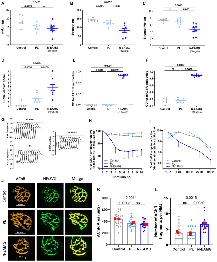
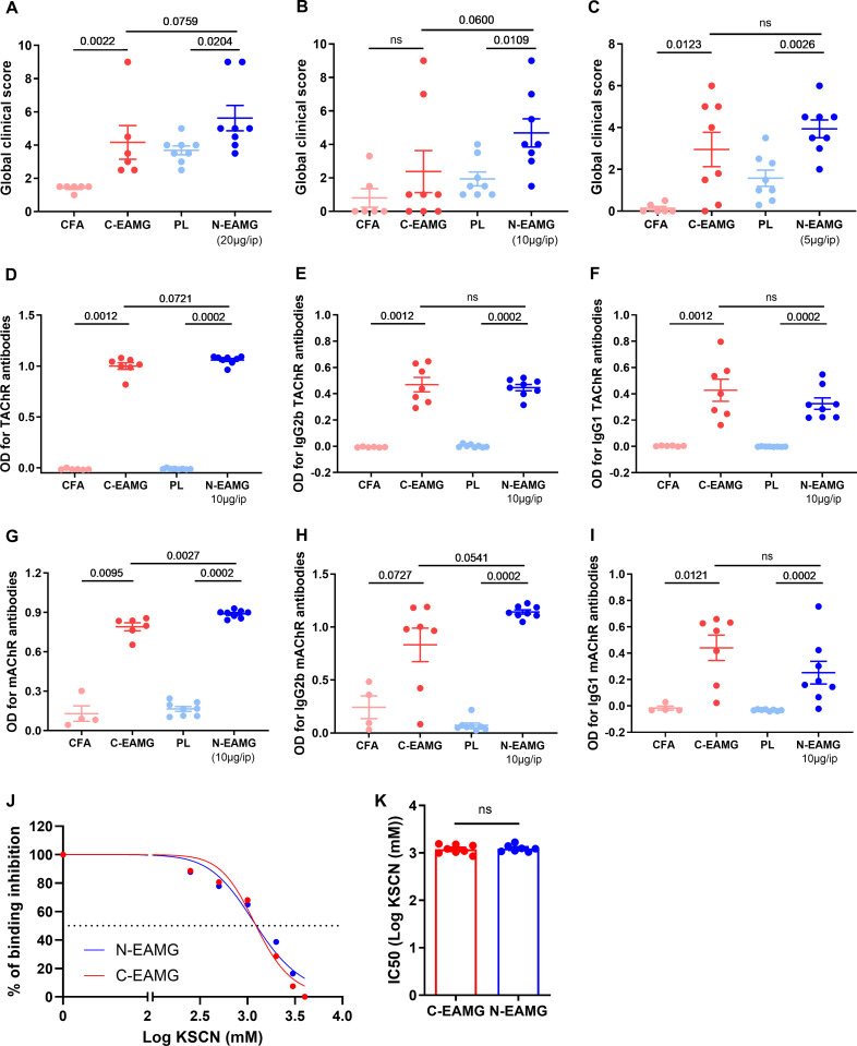
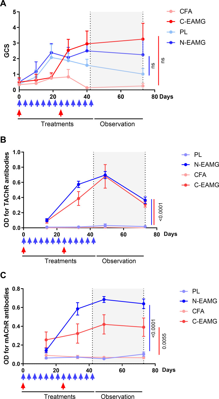
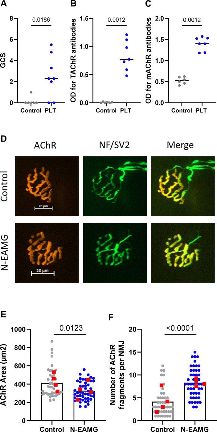
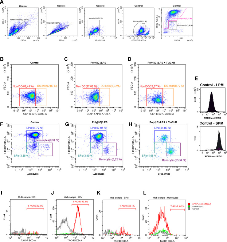
Myasthenia gravis (MG) is an autoimmune disorder primarily caused by autoantibodies that target the acetylcholine receptor (AChR) at the neuromuscular junction (NMJ). The classical experimental autoimmune myasthenia gravis (C-EAMG) mouse model has long been used by immunizing mice with acetylcholine receptor from Torpedo fish (T-AChR), combined with complete Freund's adjuvant (CFA). This mixture is administered via subcutaneous injections into the hind footpads and back, but CFA often causes strong inflammatory reactions, including lesions at the injection sites. Our objective was to develop a new EAMG model (N-EAMG) that is more compliant with animal welfare. C57Bl/6 mice were immunized twice weekly by intraperitoneal (i.p.) injection of T-AChR with a poly(I:C) and lipopolysaccharide (LPS) adjuvant mix. Control mice were injected with either physiological saline or the adjuvant mix alone. Various doses and injection schedules were tested, and the new model was compared with C-EAMG. Clinical symptoms were scored, antibody subtypes against T-AChR and mouse AChR were measured, and NMJ morphology and functionality were evaluated. We demonstrate that the N-EAMG model is at least as effective as the C-EAMG model. Moreover, similar to the C-EAMG model, the N-EAMG model is characterized by the production of T-AChR and m-AChR antibodies. This model also exhibited alterations in transmission at the NMJ due to antibody attack, resulting in a decrease in AChR surface area and increased AChR fragmentation. Symptoms were similar in both models but appeared more rapidly in the N-EAMG model. In addition, investigating the sensitization mechanism, we showed that i.p. injections of T-AChR with the poly(I:C)/LPS adjuvant mix, led to the recruitment in monocytes and changes in the two peritoneal macrophage subpopulations that were able to phagocytose T-AChR. These observations suggest that macrophage subtypes, albeit with varying efficiency, present the T-AChR to immune cells, leading to a specific immune response and the development of anti-AChR antibodies. In conclusion, our results demonstrate that this novel EAMG model is as effective as the C-EAMG model and offers several advantages. In particular, this model is more suitable for animal welfare and can replace the classical model in preclinical and fundamental research.

Keywords: LPS; adjuvant; autoimmunity; experimental model; myasthenia gravis; poly(I:C); refinement.


Conflict of interest statement

The authors declare that the research was conducted in the absence of any commercial or financial relationships that could be construed as a potential conflict of interest.

Figures

Figure 1
Figure 1
Induction of MG symptoms with i.p. injections of T-AChR: Initial proof-of-concept. C57BL/6 mice (n=6–8 per group) were i.p. injected twice weekly with physiological water (control), a poly(I:C)/LPS (PL) adjuvant mix, or a PL adjuvant mix with T-AChR 10 µg (N-EAMG). Clinical evaluations were performed after 6 weeks. (A) Mice were weighed. (B) Muscle strength was measured using a grip-test apparatus after exercise on a treadmill. (C) Muscle strength was normalized to mouse weight. (D) GCS for each mouse was calculated based on weight loss, grip test, and inverted grid test. (E, F) ELISA for anti-T-AChR and anti-m-ACHR antibodies detected with a global anti-IgG biotinylated antibody was performed on the serum collected at the end of the experiment. The integrity of neurotransmission and NMJ morphology were analyzed. Two mice with grip strength values close to the means of their respective groups were selected for these analyses. (G) CMAP was recorded in the TA muscles during a train of 10 sciatic nerve stimulations at 10 Hz. Representative traces of CMAP in control, PL, and N-EAMG mice. (H) Quantitative analysis of the CMAP amplitude after 10 Hz stimulation, expressed as a percentage of the first CMAP amplitude. (I) Quantitative analysis of the amplitude of the 10th CMAP after sciatic nerve stimulation at 1, 5, 10, 20, and 40 Hz. The data are expressed relative to the amplitude of the first CMAP of the train. (J) Representative images of isolated TA muscle fibers. Muscles were stained with α-bungarotoxin for detection of AChR (red) and antibodies directed against NF and SV2 (green) for the labeling of nerve terminals (scale bar = 20 µm). The number of AChR fragments was quantified from image stacks (control: 3 fragments, PL: 6 fragments, and N-EAMG: 7 fragments). (K) Quantitative analysis of the AChR-labeled area. (L) Number of AChR fragments isolated per NMJ. Each round point corresponds to one NMJ and each red square point corresponds to the mean value for each mouse. (A–F, K, L) P-values were assessed using the Mann-Whitney U test and indicated when p<0.1 or ns, not significant. (H, I) The data shown are the mean ± SEM (n=2; no statistical analyses were performed with such a small sample size).
Figure 2
Figure 2
Comparison of the N-EAMG model to the C-EAMG model for MG symptoms. C57BL/6 mice (n=6–8 per group) were used in three experiments with different doses of T-AChR per injection. Mice were i.p. injected twice weekly with a poly(I:C)/LPS (PL) adjuvant mix or a LP adjuvant mix with T-AChR (N-EAMG) 20 (A), 10 (B), or 5 µg (C) per injection. For the C-EAMG model, mice were immunized with CFA/T-AChR (30 µg, C-EAMG) or just CFA on day 0 and boosted after 3–4 weeks. (A–C) Clinical evaluations were performed after 6 weeks, and GCS for each mouse was calculated considering weight loss, strength, and inverted grid test results. Details of the mouse weight and strength are shown in Supplementary Figure 2 . (D–F) Anti-T-AChR antibodies were measured using ELISA and detected using anti-mouse IgG (D), IgG2b (E), and IgG1 (F) antibodies. (G–I) Anti-m-AChR antibodies were measured using ELISA and detected using anti-mouse IgG (G), IgG2b (H), and IgG1 (I) antibodies. (J, K) The relative affinity index of anti-T-AChR IgG antibodies was determined using KSCN thiocyanate, as detailed in the methods section. The binding inhibition curves represent the mean of the mice in each group (J). The half-maximal concentration of KSCN (IC50) was required to inhibit the binding of anti-AChR antibodies in the C- and N-EAMG models (K). P-values were assessed using the Mann-Whitney test to compare CFA and C-EAMG, PL and N-EAMG, C-EAMG and N-EAMG, and indicated when p<0.1 or ns, not significant.
Figure 3
Figure 3
Comparison of long-lasting effects in the N-EAMG and C-EAMG models. C57BL/6 mice (n=6-8 per group) were used in the N-EAMG and C-EAMG models. For the N-EAMG model, mice were i.p. injected twice weekly with a Poly(I:C)/LPS (PL) adjuvant mix or a PL adjuvant mix with T-AChR 10 μg (N-EAMG). Mice were injected for 6 weeks, then injections were stopped and mice were observed up to 10 weeks. For the C-EAMG model, mice were immunized with CFA/T-AChR (30µg, C-EAMG) or just CFA at day 0 and boosted after 22 days. After the boost, mice were observed up to 10 weeks. (A) Clinical evaluations were performed regularly and the global clinical score (GCS) for each mouse was calculated considering weight loss, strength, and inverted grid test results (B, C). Anti-T-AChR (B) and m-AChR (C) antibodies were measured using ELISA and detected using anti-mouse IgG. (A-C) Two-way ANOVA with Tukey post-hoc tests were performed to compare CFA and C-EAMG or PL and N-EAMG. p values were indicated when p<0.05.
Figure 4
Figure 4
Long-lasting effects affecting the NMJ in the N-EAMG model. C57BL/6 mice (n=6–8 per group) were i.p. injected twice weekly with physiological water (control) or the poly(I:C)/LPS mix with 10 µg T-AChR per injection. The mice were injected for 4 weeks, the injections were stopped, and the mice were analyzed after 4 weeks. (A) Clinical evaluations were performed regularly, and GCS for each mouse was calculated based on weight loss, grip test, and inverted grid test. (B, C) Anti-T-AChR (B) and anti-m-AChR (C) antibodies were measured using ELISA and detected using anti-mouse IgG. (D) Representative images of isolated TA muscle fibers. Muscles were stained with α-bungarotoxin for detection of AChR (red) and antibodies directed against NF and SV2 (green) for the labeling of nerve terminals (scale bar = 20 µm). The number of AChR fragments was quantified from image stacks (control: 2 fragments, and N-EAMG: 10 fragments) (E) Quantitative analysis of the AChR-labeled area. (F) The number of AChR fragments isolated per NMJ. Each round point corresponds to one NMJ and each red square point corresponds to the mean value for each mouse. P-values were assessed using the Mann-Whitney test and indicated when p<0.05.
Figure 5
Figure 5
Effects of poly(I:C)/LPS and T-AChR injections on peritoneal mononuclear phagocytes. Flow cytometry analysis of peritoneal mononuclear phagocytes (representative labeling). C57BL/6 mice (n=2 per group) were i.p. injected with physiological water, poly(I:C)/LPS (PL) or poly(I:C)/LPS containing 10 µg of T-AChR-labeled with Texas-Red. Peritoneal cells were harvested after 6 h and labeled with LIVE/DEAD™ (LD34960D) and the following antibodies: anti-CD19-APC, anti-CD3-APC, anti-CD11b-PE-Cy7, anti-CD11c-AF700, anti-Ly6C-BV605, anti-F4/80-eF480, and anti-MHC Class II-FITC. (A) Gating strategy used to identify peritoneal, singlet, living, and lineage (CD19/CD3)-negative cells, excluding eosinophils. (B–D) Gating strategy for distinguishing DC from non-DC mononuclear phagocytes. (E–H) Monocytes, SPM, and LPM were identified based on F4/80 and Ly6C labeling and the level of MHC Class II expression (E). (I–L) Detection of Texas-red labeled T-AChR in DC (I), SPM (J), LPM (K), and monocytes (L).

Similar articles

References

    1. Berrih-Aknin S, Le Panse R. Myasthenia gravis: A comprehensive review of immune dysregulation and etiological mechanisms. J Autoimmun. (2014) 52:90–100. doi: 10.1016/j.jaut.2013.12.011 - DOI - PubMed
    1. Fuchs S, Nevo D, Tarrab-Hazdai R, Yaar I. Strain differences in the autoimmune response of mice to acetylcholine receptors. Nature. (1976) 263:329–30. doi: 10.1038/263329a0 - DOI - PubMed
    1. Tuzun E, Berrih-Aknin S, Brenner T, Kusner LL, Le Panse R, Yang H, et al. . Guidelines for standard preclinical experiments in the mouse model of myasthenia gravis induced by acetylcholine receptor immunization. Exp Neurol. (2015). 270:11–7. doi: 10.1016/j.expneurol.2015.02.009 - DOI - PubMed
    1. Billiau A, Matthys P. Modes of action of Freund’s adjuvants in experimental models of autoimmune diseases. J Leukoc Biol. (2001) 70:849–60. doi: 10.1189/jlb.70.6.849 - DOI - PubMed
    1. Meinl E, Klinkert WE, Wekerle H. The thymus in myasthenia gravis. Changes typical for the human disease are absent in experimental autoimmune myasthenia gravis of the Lewis rat. Am J Pathol. (1991) 139:995–1008. - PMC - PubMed

MeSH terms

LinkOut - more resources