. 2025 May 6;13(5):e0173824.
doi: 10.1128/spectrum.01738-24. Epub 2025 Apr 15.

Nanoparticle-based biosensor integrated with multiple cross-displacement amplification for visual and rapid identification of hepatitis B virus and hepatitis C virus

Affiliations

Nanoparticle-based biosensor integrated with multiple cross-displacement amplification for visual and rapid identification of hepatitis B virus and hepatitis C virus

Hang Zhang et al. Microbiol Spectr. .

Abstract

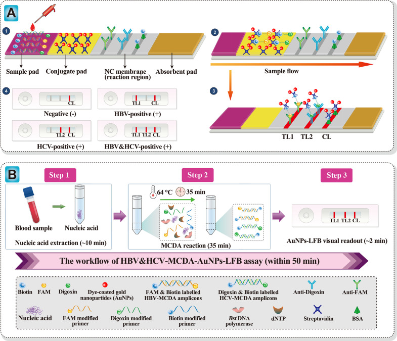
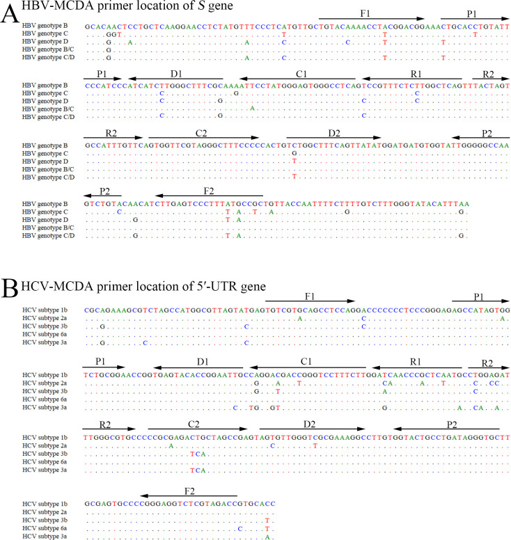
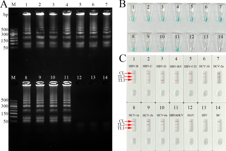
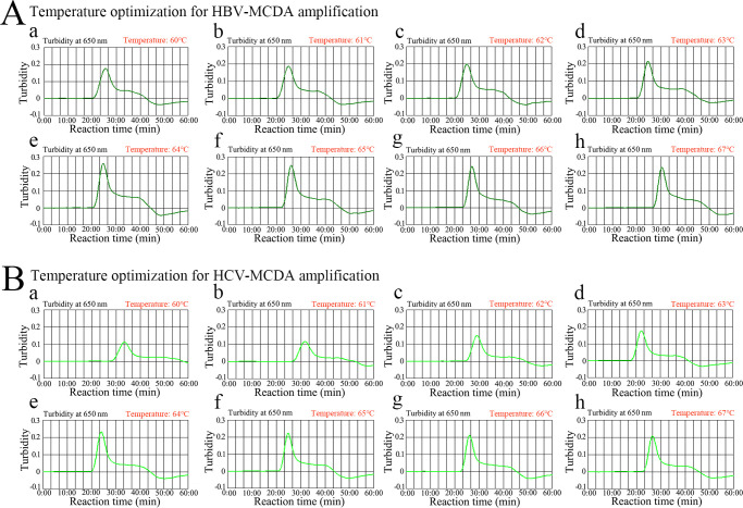
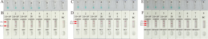
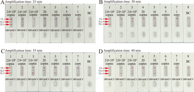
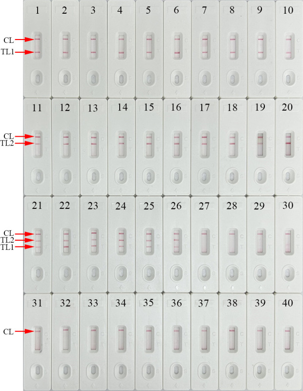
Infection with hepatitis B virus (HBV) and hepatitis C virus (HCV) is a major contributor to liver-related morbidity and mortality worldwide. An accurate and rapid point-of-care (POC) diagnostic approach is the gateway for effective treatment and control of these infections. Here, for the first time, we integrated isothermal multiple cross-displacement amplification (MCDA) with a gold nanoparticle-based lateral flow biosensor (AuNPs-LFB) to successfully develop a novel HBV&HCV-MCDA-AuNPs-LFB assay for simultaneous accurate, sensitive, rapid, inexpensive, and visual identification of HBV and HCV agents. The two unique sets of MCDA degenerate primers were successfully designed targeting the S and 5' untranslated region (5'-UTR) genes from the major HBV genotypes (B, C, D, B/C recombinant, and C/D recombinant) and HCV subtypes in China (1b, 2a, 3a, 3b, and 6a), respectively. The optimal conditions for the MCDA reaction were confirmed to be 64°C for 35 min. The MCDA products were decoded visually using the AuNPs-LFB platform, which was devised for analyzing three targets, including HBV-MCDA, HCV-MCDA amplicons, and a chromatography control. The whole detection procedure, including rapid nucleic acid extraction (~10 min), MCDA reaction (35 min), and AuNPs-LFB interpretation (~2 min), can be completed within 50 min. The HBV&HCV-MCDA-AuNPs-LFB assay can detect the target genes (HBV-S and HCV-5'-UTR) with as low as 10 copies of gene-containing plasmid template per test and does not cross-react with other pathogens. Therefore, our preliminary results indicated that the HBV&HCV-MCDA-AuNPs-LFB assay developed in this study can potentially serve as a useful POC diagnostic tool for the identification of HBV and HCV infections.IMPORTANCEHepatitis B virus (HBV) and hepatitis C virus (HCV) infections have been regarded by the World Health Organization as major threats to human health, especially in low- and middle-income regions. Underdiagnosis of HBV/HCV is a particular challenge for achieving the World Health Organization's goal of eliminating HBV and HCV infections by 2030. Here, for the first time, we integrated isothermal multiple cross-displacement amplification (MCDA) with a gold nanoparticle-based lateral flow biosensor (AuNPs-LFB) to successfully develop a novel HBV&HCV-MCDA-AuNPs-LFB assay for simultaneous accurate, sensitive, rapid, inexpensive, and visual identification and differentiation of HBV and HCV agents.

Keywords: biosensor; hepatitis B virus; hepatitis C virus; lateral flow platform; multiple cross displacement amplification.


Conflict of interest statement

The authors declare no conflict of interest.

Figures

Fig 1
Fig 1
Schematic diagram of HBV&HCV-MCDA-AuNPs-LFB assay′s principle. (A) Schematic diagram of AuNPs-LFB principles for the interpretation of HBV&HCV-MCDA products. ❶ Aliquots of 1 µL of HBV&HCV-MCDA products and 100 µL of running buffer (100 mM phosphate buffered saline (PBS), 1% Tween 20, pH 7.4) were simultaneously dripped onto the sample pad region of the biosensor. ❷ The running buffer-containing HBV&HCV-MCDA products moved forward onto the conjugate pad and NC membrane (reaction region) with capillary action. The crimson dye streptavidin-coated gold nanoparticles (streptavidin-AuNPs) are rehydrated and fixed with FAM/biotin-labeled HBV-MCDA products or digoxin/biotin-labeled HCV-MCDA products. ❸ In the reaction region, the FAM/biotin-labeled HBV-MCDA amplicons are arrested by anti-FAM at TL1. Digoxin/biotin-labeled HCV-MCDA amplicons are arrested by anti-digoxin at TL2. Excess dye-coated streptavidin-AuNPs are captured by biotinylated bovine serum albumin (biotin-BSA) at the CL region. ❹ Interpretation of the HBV&HCV-MCDA-AuNPs-LFB assay. HBV-positive results are indicated by CL and TL1 bands on the biosensor. HCV-positive results are indicated by CL and TL2 bands on the biosensor. Both HBV and HCV positive results are indicated by TL1, TL2, and CL bands on the AuNPs-LFB. Negative results are indicated when only the CL band appears on the biosensor. (B) Workflow of the HBV&HCV-MCDA-AuNPs-LFB assay. The HBV&HCV-MCDA-AuNPs-LFB assay’s workflow comprises the following closely linked steps: rapid nucleic acid extraction (step 1), MCDA reaction (step 2), and AuNPs-LFB visual readout (step 3). The whole diagnostic procedure can be completed within 50 min.
Fig 2
Fig 2
Nucleotide sequences and location of the HBV-S and HCV 5′-UTR genes used to design the HBV&HCV-MCDA primers. In the MCDA reaction, the isothermal amplification of specific nucleic acid sequences is achieved by employing a set of 10 primers spanning 10 distinct regions of the target fragment, which are designated as displacement primers F1 and F2, cross primers CP1 (C1 and P1 go together) and CP2 (C2 and P2 go together), and amplification primers (C1, C2, D1, D2, R1, and R2). (A) The nucleotide sequences of the S gene from five dominant HBV genotypes in China (B, C, D, recombinant B/C, and recombinant C/D) were aligned by DNASTAR software, and the HBV-MCDA primer sequences were marked with arrows. (B) The nucleotide sequences of the 5′-UTR gene from five dominant HCV subtypes in China (1b, 2a, 3a, 3b, and 6a) were aligned by DNASTAR software, and the conserved sequences were applied for HCV-MCDA primers. Right arrows and left arrows indicated the sense and complementary sequences which were used in this study, respectively.
Fig 3
Fig 3
Confirmation and detection of HBV-, HCV-, and HBV&HCV-MCDA products. (A) 2% agarose gel electrophoresis, (B) visual indicator (MG), (C) AuNPs-LFB biosensor. Templates of 1–14 were HBV-B plasmid, HBV-C plasmid, HBV-D plasmid, HBV-B/C plasmid, HBV-C/D plasmid, HCV-1b plasmid, HCV-2a plasmid, HCV-3a plasmid, HCV-3b plasmid, HCV-6a plasmid, HBV&HCV plasmid, HAV, and HIV, respectively. For agarose gel electrophoresis detection, the agarose gel presented ladder-like bands indicating a positive outcome, whereas the lack of bands indicated a negative result. For visual MG analysis, changing the reaction mixture to light green suggested a positive outcome, whereas colorless mixtures indicated a negative result. For AuNPs-LFB identification, both the CL and TL1 simultaneously appeared on the AuNPs-LFB strip, demonstrating an HBV-positive result. Both the CL and TL2 simultaneously turned red on the biosensor, indicating an HCV-positive outcome. For a negative outcome, only CL was present on the AuNPs-LFB strips.
Fig 4
Fig 4
Temperature optimization for HBV- and HCV-MCDA amplification. The MCDA reactions for detection of HBV (A) and HCV (B) were monitored using real-time turbidity, and their corresponding amplicon curves were shown as graphs. A turbidity >0.1 indicated a positive result. Eight kinetic graphs (a–h) were obtained at different temperatures (60°C–67°C, 1°C increments) with 1 × 10⁴ target gene copies. Graphs e (64°C) to h (67°C) in A showed robust amplification. Graphs from e (64°C) to f (65°C) in B showed robust amplification.
Fig 5
Fig 5
Sensitivity analysis of HBV&HCV-MCDA-AuNPs-LFB assay with serial nucleic acid template dilutions. Serial dilutions of HBV-B and HCV-1b plasmids were used as templates, and distilled water was used as the BC. Results were simultaneously analyzed by visual reagent MG and AuNPs-LFB biosensor. (A, B) Sensitivity analysis of HBV-MCDA reaction. Tubes A1–A8 (biosensor B1–B8) represent the HBV-S plasmid amounts of 2.0 × 10⁴ copies, 2.0 × 10³ copies, 2.0 × 10² copies, 20 copies, 10 copies, 5 copies, 1 copy per reaction, and blank control, respectively. The LoD of HBV-MCDA assay was 10 copies per reaction. (C, D) Sensitivity analysis of HCV-MCDA reaction. Tubes C1–C8 (biosensor D1–D8) represent the HCV-5′-UTR plasmid amounts of 2.0 × 10⁴ copies, 2.0 × 10³ copies, 2.0 × 10² copies, 20 copies, 10 copies, 5 copies, 1 copy per reaction, and blank control, respectively. The LoD of the HCV-MCDA assay was 10 copies per reaction. (E, F) Sensitivity analysis of HBV&HCV-MCDA reaction. Tubes E1–E8 (biosensor F1–F8) represent the HBV&HCV plasmid amounts of 2.0 × 10⁴ copies, 2.0 × 10³ copies, 2.0 × 10² copies, 20 copies, 10 copies, 5 copies, 1 copy per reaction, and blank control, respectively. The LoD of HBV&HCV-MCDA assay was 10 copies per reaction. For visual MG analysis, changing the reaction mixture to light green suggested a positive outcome, whereas colorless mixtures indicated a negative result. For AuNPs-LFB identification, both the CL and TL1 simultaneously appeared on the AuNPs-LFB strip, demonstrating an HBV-positive result. Both the CL and TL2 simultaneously turned red on the biosensor, indicating an HCV-positive outcome. For a negative outcome, only CL was present on the AuNPs-LFB strips.
Fig 6
Fig 6
Amplification time optimization for HBV&HCV-MCDA-AuNPs-LFB assay. Different amplification times (A, 25 min; B, 30 min; C, 35 min; D, 40 min) were evaluated at optimal reaction temperature (64℃). Biosensors 1–8 represent HBV-B and HCV-1b nucleic acid template levels of 2.0 × 10⁴ copies, 2.0 × 10³ copies, 2.0 × 10² copies, 20 copies, 10 copies, 5 copies, 1 copy, and blank control (distilled water), respectively. HBV&HCV-MCDA amplicons were analyzed using AuNPs-LFB biosensor. The optimal LoD occurred with a 35 min amplification time (C).
Fig 7
Fig 7
Specificity analysis of HBV&HCV-MCDA-AuNPs-LFB assay with different strains. The MCDA reactions were performed using different nucleic acid templates, and each of the amplification products was analyzed through visual AuNPs-LFB biosensor. Biosensor 1–5, HBV genotype B plasmid, HBV genotype C plasmid, HBV genotype D plasmid, HBV genotype B/C plasmid, HBV genotype C/D plasmid, respectively; biosensor 6–10, HBV strains (clinical samples); biosensor 11–15, HCV subtype 1b plasmid, HCV subtype 2a plasmid, HCV subtype 3b plasmid, HCV subtype 6a plasmid, HCV subtype 3a plasmid, respectively; biosensor 16–20, HCV strains (clinical samples); biosensor 21, HBV&HCV plasmids; biosensor 22–26, HBV&HCV strains (clinical samples); biosensor 27, herpes simplex virus; biosensor 28, parainfluenza virus; biosensor 29, HAV; biosensor 30, coxsackievirus CAV16; biosensor 31, human enterovirus EV71; biosensor 32, influenza A virus; biosensor 33, influenza B virus; biosensor 34, human papilloma virus; biosensor 35, HIV; biosensor 36, Mycobacterium leprae; biosensor 37, Mycobacterium tuberculosis; biosensor 38, Haemophilus influenzae; biosensor 39, Cryptococcus neoformans; biosensor 40, blank control (distilled water).

Similar articles

References

    1. Said ZNA, El-Sayed MH. 2022. Challenge of managing hepatitis B virus and hepatitis C virus infections in resource-limited settings. World J Hepatol 14:1333–1343. doi:10.4254/wjh.v14.i7.1333 - DOI - PMC - PubMed
    1. Alberts CJ, Clifford GM, Georges D, Negro F, Lesi OA, Hutin YJ-F, de Martel C. 2022. Worldwide prevalence of hepatitis B virus and hepatitis C virus among patients with cirrhosis at country, region, and global levels: a systematic review. Lancet Gastroenterol Hepatol 7:724–735. doi:10.1016/S2468-1253(22)00050-4 - DOI - PMC - PubMed
    1. Cui F, Blach S, Manzengo Mingiedi C, Gonzalez MA, Sabry Alaama A, Mozalevskis A, Séguy N, Rewari BB, Chan P-L, Le L, Doherty M, Luhmann N, Easterbrook P, Dirac M, de Martel C, Nayagam S, Hallett TB, Vickerman P, Razavi H, Lesi O, Low-beer D. 2023. Global reporting of progress towards elimination of hepatitis B and hepatitis C. Lancet Gastroenterol Hepatol 8:332–342. doi:10.1016/S2468-1253(22)00386-7 - DOI - PubMed
    1. Thomas DL. 2019. Global elimination of chronic hepatitis. N Engl J Med 380:2041–2050. doi:10.1056/NEJMra1810477 - DOI - PubMed
    1. Cox AL, El-Sayed MH, Kao JH, Lazarus JV, Lemoine M, Lok AS, Zoulim F. 2020. Progress towards elimination goals for viral hepatitis. Nat Rev Gastroenterol Hepatol 17:533–542. doi:10.1038/s41575-020-0332-6 - DOI - PMC - PubMed

MeSH terms

LinkOut - more resources