. 2025 May 20;99(5):e0210524.
doi: 10.1128/jvi.02105-24. Epub 2025 Apr 15.

A monomeric envelope glycoprotein cytoplasmic tail is sufficient for HIV-1 Gag lattice trapping and incorporation

Affiliations

A monomeric envelope glycoprotein cytoplasmic tail is sufficient for HIV-1 Gag lattice trapping and incorporation

Nicholas S Groves et al. J Virol. .

Abstract

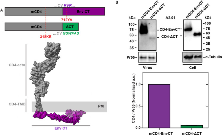
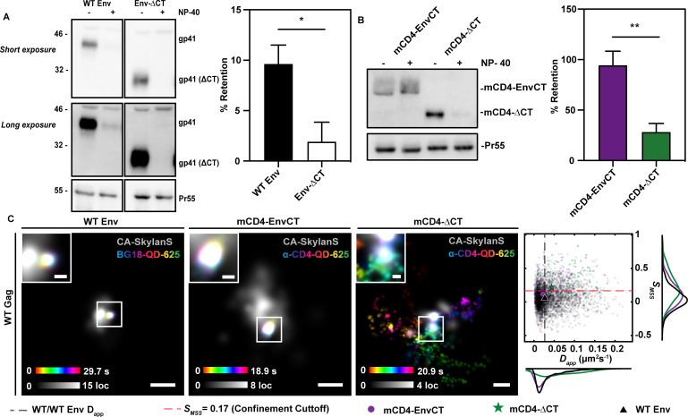
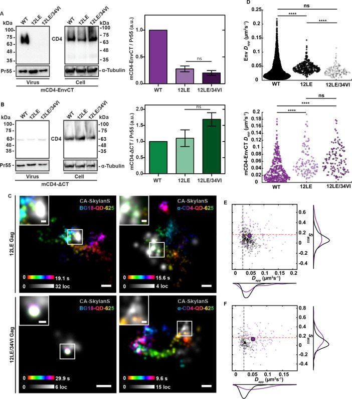
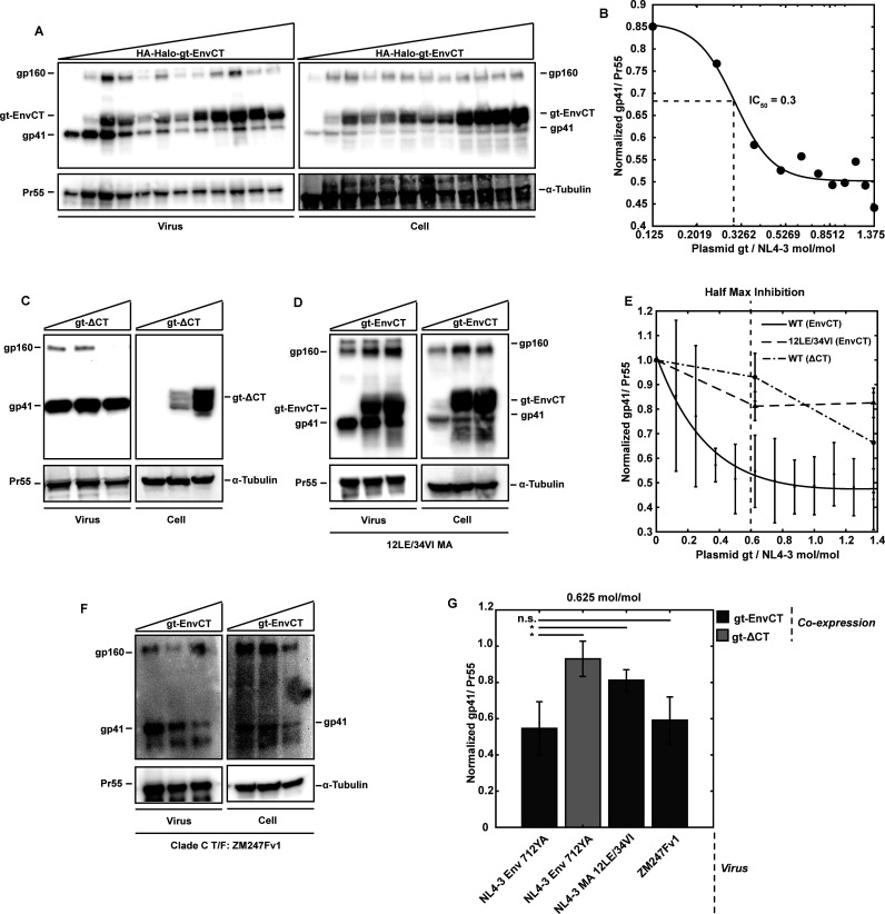
To become infectious, assembling enveloped viruses must acquire viral glycoproteins to mediate downstream infection events. Human immunodeficiency virus-1 (HIV-1) envelope glycoproteins (Env) are well characterized to function as trimers for membrane fusion and entry; however, we sought to understand whether the trimeric structure of Env is required for incorporation into virus particles. Using superresolution live-cell imaging and biochemical assays, we demonstrate that a monomeric receptor chimera containing the Env cytoplasmic tail (Env-CT), known to regulate Env incorporation, is sufficient for lattice trapping and incorporation into virus assembly sites. We also demonstrate that these Env-CT monomers can restrict the incorporation of native Env trimers, competing for an apparently limited number of interaction sites in each assembling particle. Furthermore, this monomeric construct can restrict the incorporation of Env glycoproteins from an evolutionarily distant HIV-1 primary isolate. Our findings support a model where a monomeric Env-CT mediates Env incorporation, with this mechanism of Env incorporation being conserved between distant clades of HIV-1.IMPORTANCETo combat the prevalence of HIV-1 and antiviral resistance, new classes of antivirals are needed. An attractive target for new classes includes virus assembly because released virus particles unable to obtain Env glycoproteins are non-infectious and unable to propagate HIV-1 infection. One requisite to the development of an antiviral targeting Gag-Env coalescence is the need to define the functional units constituting this molecular interface. Although Env functions as an obligatory trimer for virus entry, we demonstrate that a monomeric Env-CT is sufficient for Env incorporation into HIV-1 particles. Monomeric Env-CT displayed saturability in viral lattices and the ability to compete with native Env trimers for particle incorporation. These results suggest a less complex Env-CT structure mediates virus incorporation and that Env-CT mimetics could yield broad competitive activity against HIV-1 infection.

Keywords: Env; Gag; HIV-1; biochemistry; glycoprotein incorporation; single-particle tracking; virus assembly.


Conflict of interest statement

The authors declare no conflict of interest

Figures

Fig 1
Fig 1
Monomeric HIV-1 Env cytoplasmic tail chimeras are incorporated into virus particles in an Env-CT-dependent manner. (A) Genetic design of a monomeric CD4-EnvCT 712YA chimera and hypothetical domain organization (Env-CT: PDBID: 5VWL, CD4: PDBID:1WIP). The GSWPAS peptide motif is the epitope for the anti-EnvCT antibody Chessie8 and is appended to the mCD4-∆CT construct to enable comparative Western blotting. (B) A2.01 T-cell lines stably expressing mCD4-EnvCT or mCD4-∆CT were transduced with NL4-3 virus (∆pol/∆vif/∆vpr/∆env). After 48 hours, virus and cell samples were analyzed by Western blot using anti-CD4, anti-p24, and anti-tubulin antibodies (top). The A2.01 T-cell line is null for native CD4. HIV-1 particle incorporation of mCD4-EnvCT in the A2.01 T-cell line is strongly dependent on the Env-CT, suggesting that a monomeric Env-CT is sufficient for Gag-lattice enrichment (mCD4-EnvCT /mCD4-∆CT = 16.7-fold).
Fig 2
Fig 2
A monomeric Env-CT chimera is retained in the immature HIV-1 Gag lattice. (A, B) HEK293T cells were co-transfected with pNL4-3 ΔpolΔenv and mCD4-EnvCT or mCD4-ΔCT chimera plasmids. VLPs containing HIV-1 WT Env or EnvΔCT were produced using pNL4-3 Δpol or pNL4-3 Δpol CTdel144. After 48 hours, the virus was harvested, and membranes were stripped using NP-40 detergent. Samples were immunoblotted for Env (10E8v4/2F5), CD4 (ab133616), and Pr55 (HIV-Ig). Approximately 10% of WT Env is shown to co-sediment with Gag (Pr55) after detergent treatment, whereas Env-ΔCT trimers are stripped from immature VLPs (A) (P = 0.025, n = 4). Similarly, mCD4-EnvCT monomers co-sediment with Gag while mCD4-ΔCT is largely stripped from immature VLPs. (B) (P = 0.0066, n = 4). (C) WT COS7 cells (left) or COS7 cells stably expressing mCD4-EnvCT (middle)/mCD4-∆CT (right) were infected with NL4-3 virus (∆pol/∆vif/∆vprenv/∆nef::anti-CA-SkylanS). After 38–42 hours, cells were labeled with anti-Env-QD625 or anti-CD4-QD625 conjugates and imaged using TIRF microscopy at 37°C and 5% CO2. Gag was detected by coexpression and imaging of anti-CA nanobody fused to the photoswitchable SkylanS fluorescent protein. Time projections of individual single-particle tracks (color bar) proximal to superresolved HIV-1 assembly sites (gray). Individual tracks display confined or Brownian mobility dependent on the presence or absence of an Env-CT, respectively (scale bars = 200 nm, insets = 50 nm). Gray scale bars indicate Gag localization (loc) density over the time of acquisition, and color bars indicate particle position over the observation period. Confined tracks (mCD4-EnvCT, WT Env) appear white and centered in the insets. Scatter histograms of diffusion parameters: Dapp and SMSS for WT Env and chimera monomers with respect to WT Gag lattice assembly sites indicate a significant increase in mobility for mCD4-ΔCT as compared to mCD4-EnvCT and WT Env (mCD4-EnvCT: Dapp = 0.022 ± 5.7×10⁻⁴ µm2s−1, SMSS = 0.12 ± 0.18; mCD4-ΔCT: Dapp = 0.044 ± 1.6×10⁻³ µm2s−1, SMSS = 0.22 ± 0.22; WT Env: Dapp = 0.027 ± 1.9×10⁻⁴ µm2s−1, SMSS = 0.056 ± 0.16; mCD4-EnvCT, WT Env: PDapp = 2.7 × 10⁻⁴, PSMSS = 1.2 × 10⁻¹³). The red dashed line, SMSS >0.17, indicates Brownian motion. The black dashed line represents the mean Dapp for WT Env. All errors represent SD.
Fig 3
Fig 3
A monomeric Env-CT is sensitive to perturbation of the Gag MA domain. (A, B) A2.01 T-cells stably expressing mCD4 ± EnvCT were infected with a single-round infectious virus possessing the indicated Gag genotypes (NL4-3-∆pol/∆vif/∆vpr/∆env). After ~48 hours, virus supernatants and cells were harvested and subjected to Western blotting. (A) The 12LE and 12LE/34VI MA lattice are defective for mCD4-EnvCT incorporation. (B) mCD4-∆CT chimeras are insensitive to the 12LE and 12LE/34VI MA mutant lattice and passively incorporate regardless of the MA genotype. (C) COS7 cells stably expressing mCD4-EnvCT were infected with a single-round infectious virus possessing the indicated Gag genotypes (NL4-3-∆pol/∆vif/∆vprenv/∆nef::anti-CA-SkylanS). After 38–42 hours, native Env trimers were labeled with anti-Env (BG18)-QD625 (left panels) or mCD4-EnvCT molecules were labeled with anti-CD4-QD-625 conjugates (right panels) and imaged by TIRF microscopy at 37°C with 5% CO2. Reconstructions of Gag assembly sites were performed by single molecule sampling of expressed anti-CA nanobody fused to the photoswitchable SkylanS fluorescent protein. Representative time projections of single molecule trajectories for WT Env and mCD4-EnvCT diffusion in 12LE and 12LE/34VI MA lattices (mCD4-EnvCT: Dapp = 0.049 ± 1.3×10⁻³ µm2s−1, SMSS = 0.15 ± 0.19; WT Env: Dapp = 0.043 ± 3.9×10⁻⁴ µm2s−1, SMSS = 0.067 ± 0.12, PDapp = 1.1 × 10⁻³, PSMSS = 3.4×10⁻⁸). Diffusion in 12LE/34VI lattices (mCD4-EnvCT: Dapp = 0.056 ± 1.4×10⁻³ µm2s−1, SMSS = 0.14 ± 0.17; WT Env: Dapp = 0.029 ± 6.2×10⁻⁴ µm2s−1, SMSS = 0.058 ± 0.14, PDapp = 3.6×10⁻¹⁷, PSMSS = 7.5×10⁻⁵). Scale bars = 200 nm, inset = 50 nm. (D) Apparent diffusion coefficients for WT Env (top panel) and mCD4-EnvCT monomer (bottom panel) for all virus assembly sites measured. WT Env (BG18-QD-625) confinement is defective in the 12LE MA lattice; however, WT Env becomes diffusionally confined with the 12LE/34VI MA lattice rescue mutation (WT MA, 12LE: PDapp = 2.9×10⁻²³; 12LE, 12LE/34VI: PDapp = 3.6×10⁻¹⁷; WT MA, 12LE/34VI: PDapp = 0.16). Diffusional confinement is not rescued in the 12LE/34VI lattice for mCD4-EnvCT monomers (WT MA, 12LE: PDapp = 3.7×10⁻¹⁵; 12LE, 12LE/34VI: PDapp = 0.14; WT MA, 12LE/34VI: PDapp = 8.5×10⁻¹⁹). (E, F) mCD4-EnvCT and WT Env have differing diffusion modalities in both the 12LE and 12LE/34VI mutant MA lattices, shown by SMSS and Dapp in scatter-histograms. The red dashed line indicates SMSS >0.17, Brownian motion cutoff. The black dashed line indicates the mean Dapp for WT Env/MA diffusion. Black triangles are mean WT Env and magenta circles are mean mCD4-EnvCT values. All errors represent S.D.
Fig 4
Fig 4
Monomeric Env-CT competes with native Env trimers for virion incorporation. Env incorporation competition experiments were performed by transfecting HEK293T cells with plasmids for single-round infectious HIV-1 (NL4-3- or ZM247Fv2-∆pol/∆vif/∆vpr) and HA-Halo-gt-EnvCT(712YA) chimera. (A) Representative western blots of VLPs and cell lysates for competitive inhibition experiments using Env 712YA with increasing plasmid concentrations of monomeric HA-Halo-gt-EnvCT(712YA). (B) A dose-response curve was estimated using a sigmoidal fit (IC50 = 0.3 molgt/molNL4-3; mean square error = 1.2 × 10⁻³). (C) Western blots demonstrate that HA-Halo-gt-∆CT does not compete with Env 712YA with increasing plasmid ratios (0, 0.625, and 1.375 molgt/molNL4-3). (D) Western blots showing monomeric HA-Halo-gt-EnvCT cannot inhibit WT Env incorporation in the 12LE/34VI mutant MA lattice. For all western blots, gp41 (Env 712YA) and HA-Halo-gt-EnvCT were detected with anti-EnvCT (Chessie8) mAb, Gag was detected with anti-p24 (183-H12-5C) mAb, and the HA epitope with anti-HA (H17-L2) mAb. (E) Double exponential loss curve of the competitive inhibition of 712YA Env with half-maximal inhibition occurring at 0.6 ± 0.1 gp41/Pr55 (vertical dashed line represents half-maximum of inhibition; root mean square error of fit = 0.04). (F) Clade C transmitted founder virus Env competition for virus assembly sites is observed with expression of HA-Halo-gt chimera possessing the Clade B (NL4-3) Env-CT. (G) Comparisons of the mean gp41/Pr55 ratio between all assays performed at 0.625 molgt/molNL4-3 plasmid ratios (MA-WT/Env-712A versus MA-WT/gt-∆CT: P = 0.02; MA-WT/Env-712YA versus MA-12LE-34VI/gt-EnvCT: P = 0.04; MA-WT/gt-∆CT versus MA-12LE-34VI/gt-EnvCT: P = 0.15, MA-WT/Env-712YA versus ZM247Fv1/gt-EnvCT: P = 0.69). Error bars represent SD.

Similar articles

References

    1. Jouvenet N, Bieniasz PD, Simon SM. 2008. Imaging the biogenesis of individual HIV-1 virions in live cells. Nature 454:236–240. doi:10.1038/nature06998 - DOI - PMC - PubMed
    1. Briggs JAG, Wilk T, Welker R, Kräusslich HG, Fuller SD. 2003. Structural organization of authentic, mature HIV-1 virions and cores. EMBO J 22:1707–1715. doi:10.1093/emboj/cdg143 - DOI - PMC - PubMed
    1. Willey RL, Bonifacino JS, Potts BJ, Martin MA, Klausner RD. 1988. Biosynthesis, cleavage, and degradation of the human immunodeficiency virus 1 envelope glycoprotein gp160. Proc Natl Acad Sci USA 85:9580–9584. doi:10.1073/pnas.85.24.9580 - DOI - PMC - PubMed
    1. Moulard M, Hallenberger S, Garten W, Klenk HD. 1999. Processing and routage of HIV glycoproteins by furin to the cell surface. Virus Res 60:55–65. doi:10.1016/s0168-1702(99)00002-7 - DOI - PubMed
    1. Hendrix J, Baumgärtel V, Schrimpf W, Ivanchenko S, Digman MA, Gratton E, Kräusslich H-G, Müller B, Lamb DC. 2015. Live-cell observation of cytosolic HIV-1 assembly onset reveals RNA-interacting Gag oligomers. J Cell Biol 210:629–646. doi:10.1083/jcb.201504006 - DOI - PMC - PubMed

MeSH terms

Substances

LinkOut - more resources