Case Reports
. 2025 Sep 1;27(5):598-610.
doi: 10.4103/aja2024107. Epub 2025 Apr 15.

Recurrent spontaneous miscarriages from sperm after ABVD chemotherapy in a patient with Hodgkin's lymphoma: sperm DNA and methylation profiling

Affiliations
Case Reports

Recurrent spontaneous miscarriages from sperm after ABVD chemotherapy in a patient with Hodgkin's lymphoma: sperm DNA and methylation profiling

Gwendoline Lecuyer et al. Asian J Androl. .

Abstract

Lymphomas represent one of the most common malignant diseases in young men and an important issue is how treatments will affect their reproductive health. It has been hypothesized that chemotherapies, similarly to environmental chemicals, may alter the spermatogenic epigenome. Here, we report the genomic and epigenomic profiling of the sperm DNA from a 31-year-old Hodgkin lymphoma patient who faced recurrent spontaneous miscarriages in his couple 11-26 months after receiving chemotherapy with adriamycin, bleomycin, vinblastine, and dacarbazine (ABVD). In order to capture the potential deleterious impact of the ABVD treatment on mutational and methylation changes, we compared sperm DNA before and 26 months after chemotherapy with whole-genome sequencing (WGS) and reduced representation bisulfite sequencing (RRBS). The WGS analysis identified 403 variants following ABVD treatment, including 28 linked to genes crucial for embryogenesis. However, none were found in coding regions, indicating no impact of chemotherapy on protein function. The RRBS analysis identified 99 high-quality differentially methylated regions (hqDMRs) for which methylation status changed upon chemotherapy. Those hqDRMs were associated with 87 differentially methylated genes, among which 14 are known to be important or expressed during embryo development. While no variants were detected in coding regions, promoter regions of several genes potentially important for embryo development contained variants or displayed an altered methylated status. These might in turn modify the corresponding gene expression and thus affect their function during key stages of embryogenesis, leading to potential developmental disorders or miscarriages.

Keywords: chemotherapy; embryo loss; methylation; miscarriage; sperm DNA; whole-genome sequencing.


Conflict of interest statement

All authors declare no competing interests.

Figures

Figure 1
Figure 1
Description of patient pattern. The Hodgkin lymphoma patient cryopreserved sperm before 6 cycles of ABVD chemotherapy in 2008. The chemotherapy protocol ended in June 2009. The couple obtained four natural pregnancies between May 2010 and September 2011, but all ended as miscarriages. Semen parameters were normal in September 2011, but due to the miscarriages an assisted reproduction method (intra-uterine-insemination [IUI] of 8.4 × 10⁶ motile sperm from cryopreserved sperm straws before chemotherapy) was used and allowed to obtain a healthy child in 2013. CT: chemotherapy; ABVD: adriamycin, bleomycin, vinblastine, and dacarbazine; WGS: whole genome sequencing; RRBS: reduced representation of bisulfite sequencing; HL: Hodgkin lymphoma; GWAS: Genome Wide Association Study.
Figure 2
Figure 2
Filtering strategy to select variants and functional annotation. (a) The numbers of selected single-nucleotide polymorphisms (SNPs) and insertions/deletion variants (INDELs) at each step and for each tool are indicated. This strategy is based on (1) read coverage; (2) difference of variant frequency ≥50% between the sample before chemotherapy (pre-CT) and the sample after chemotherapy (post-CT) samples; (3) variants must be detected by at least 2 tools including VarScan; and (4) variant frequency in the general population ≤0.1%. (b) Variant annotation to genomic regions. The association to genes important for embryogenesis is indicated. (c) Variant annotation to regulatory elements and distance to transcription start site (TSS) in kilobases (kb) are indicated. TF: transcription factor; CDS: coding DNA sequence; UTR: untranslated regions; NMD: nonsense-mediated mRNA decay.
Figure 3
Figure 3
Read mapping and methylation statistics for the sample before (pre-CT) and after chemotherapy (post-CT). (a) Read mapping statistics. (b) Sample-wise numbers of CpG, CHG and CHH (H corresponds to A, T or C). (c) Sample-wise coverage distribution of CpG sites. Distribution of coverage across samples after coverage limitation (90% quantile). (d) Proportion of methylated CpG, CHG and CHH for each sample. (e) Distribution of methylated CpGs among genomic features. A minimum depth of four reads is required for each CpG. CpG methylation levels are averaged by feature to produce feature-associated methylation levels.
Figure 4
Figure 4
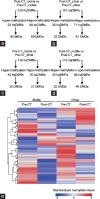
Filtration of high-quality differentially methylated regions (hqDMRs) between the sample before (pre-CT) and after chemotherapy (post-CT). Number of hypermtheliated and hypomethylated hqDMRs and related differentially methylated genes (DMGs): (a) in the post-CT_motile vs pre-CT_motile comparison; (b) in the post-CT_other vs pre-CT_other comparison; (c) in the pre-CT_motile vs pre-CT_other comparison; and (d) in the post-CT_motile vs post-CT_other comparisons. (e) A false-color heatmap of standardized methylation levels of the resulting 273 hqDMRs which have been sorted (dendrogram on the left and on the top) based on a hierarchical clustering applied to the standardized methylation level data. Each line corresponds to an hqDMR and each column to a sample. Standardized methylation levels are displayed according to a color code (bottom right) ranging from blue (hypomethylation) to red (hypermethylation).
Figure 5
Figure 5
Heatmap of the 129 hqDMRs showing altered methylation status after chemotherapy in mobile sperms (post-CT_motile). The numbers of high-quality differentially methylated regions (hqDMRs) and related differential methylated genes (DMGs) are given for each of the seven methylation patterns (P1–P7) on the right, while the overall number of hyper- and hypo-methylated hqDMRs is indicated on the left. Each line corresponds to an hqDMR and each column to a sample. Standardized methylation levels are displayed according to a color code (bottom right) ranging from blue (low methylation level) to red (high).

References

    1. Ferlay J, Soerjomataram I, Dikshit R, Eser S, Mathers C, et al. Cancer incidence and mortality worldwide:sources, methods and major patterns in GLOBOCAN 2012. Int J Cancer. 2015;136:E359–86. - PubMed
    1. National Estimates of Cancer Incidence and Mortality in Metropolitan France between 1990 and 2018 – Malignant Hematological Diseases: A Study Based on Data from the Francim Cancer Registry Network. [[Last accessed on 2024 Dec 25]]. Available from: https://www.santepubliquefrance.fr/docs/estimations-nationales-de-l-inci... .
    1. Shankland KR, Armitage JO, Hancock BW. Non-Hodgkin lymphoma. Lancet. 2012;380:848–57. - PubMed
    1. Ansell SM. Hodgkin lymphoma:diagnosis and treatment. Mayo Clin Proc. 2015;90:1574–83. - PubMed
    1. Fanale MA, Younes A. Monoclonal antibodies in the treatment of non-Hodgkin's lymphoma. Drugs. 2007;67:333–50. - PubMed

Publication types

MeSH terms

Supplementary concepts