

Optical coherence tomography reveals retinal structural abnormalities in α-synucleinopathies: insights from the Padua-CESNE cohort
- PMID: 40232370
- PMCID: PMC12208970
- DOI: 10.1007/s00702-025-02918-y
Optical coherence tomography reveals retinal structural abnormalities in α-synucleinopathies: insights from the Padua-CESNE cohort
Abstract
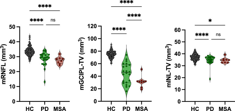
The complexity of α-synucleinopathies, namely Parkinson's disease (PD) and multiple system atrophy (MSA), calls for the adoption a multimodal approach integrating biological, morphological, and functional data. Phosphorylated α-synuclein (α-syn) detection in bodily fluids and tissues such as the skin helps provide biological characterization of the disease, but specific and accessible biomarkers are not available yet. The aim of this study was to define the role of Optical Coherence Tomography (OCT, a minimally invasive retinal imaging technique) patterns as possible biomarkers in the early stages of α-synucleinopathies, also supporting the differential diagnosis. Thirty-five (23 PD, 12 MSA), clinically, biologically and genetically characterized patients included in the PADUA-CESNE (Centro Studi per la Neurodegenerazione) cohort underwent OCT. A significant atrophy in the inferior, superior and temporal regions of the Retinal Nerve Fiber Layer (RNFL) and in the inner nuclear layer (INL) were observed in PD compared to controls, differently from MSA. Hyperreflective foci (HRF) counts were elevated across all retinal layers in all patients with PD exhibiting significantly higher numbers, suggesting microglial activation and greater retinal damage. Further research regarding OCT patterns in PD and MSA may consolidate the role of specific features, such as INL abnormalities and different HRF counts, in supporting the diagnosis and differential diagnosis in α-synucleinopathies. In light of the availability of potentially disease-modifying therapies, studies should focus on newly diagnosed patients, also undergoing thorough clinical, biological and genetic characterization.
Keywords: Biomarkers; Multiple system atrophy; Optical coherence tomography; Parkinson’s disease; α-synuclein.
© 2025. The Author(s).
Conflict of interest statement
Declarations. Conflict of interest: The authors declare no disclosures or conflicts of interest related to the manuscript's content. AA has received compensation for consultancy and speaker related activities from UCB, Bayer, Ever Pharma, Britannia, AbbVie, Zambon, Bial, Theravance Biopharma, Jazz Pharmaceuticals, Roche, Medscape; he receives research support from Horizon 2020, Italian Ministry of University and Research (MUR), Italian Ministry of Health, Next Generation EU—National Center for Gene Therapy and Drugs based on RNA Technology and National Recovery and Resilience Plan, Investment PE8 – Project Age-It: “Ageing Well in an Ageing Society”.MC has received travel grants from Zambon and receives support from Progetto di ricerca di Rilevante Interesse Nazionale – PRIN 2022 FJAXY8. All the other authors have no financial disclosure to declare. Informed consent: Written informed consent was obtained from each participant after a detailed description of the study. Informed consent for the use of biological samples was obtained from all patients. All procedures on human tissue samples were carried out in accordance with the Declaration of Helsinki.
Figures
Similar articles
-
Chronic urinary tract infections cause persistent microglial changes in a humanized ɑ-synuclein mouse model.J Parkinsons Dis. 2024 Nov;14(8):1559-1574. doi: 10.1177/1877718X241289046. Epub 2024 Dec 27. J Parkinsons Dis. 2024. PMID: 39957188
-
Spectral domain and angiography optical coherence tomography in atypical parkinsonisms and Parkinson disease: an explorative study.Parkinsonism Relat Disord. 2025 Aug;137:107932. doi: 10.1016/j.parkreldis.2025.107932. Epub 2025 Jun 16. Parkinsonism Relat Disord. 2025. PMID: 40543174
-
Neuropathological stages of neuronal, astrocytic and oligodendrocytic alpha-synuclein pathology in Parkinson's disease.Acta Neuropathol Commun. 2025 Feb 11;13(1):25. doi: 10.1186/s40478-025-01944-x. Acta Neuropathol Commun. 2025. PMID: 39934841 Free PMC article.
-
Optical coherence tomography (OCT) for detection of macular oedema in patients with diabetic retinopathy.Cochrane Database Syst Rev. 2011 Jul 6;(7):CD008081. doi: 10.1002/14651858.CD008081.pub2. Cochrane Database Syst Rev. 2011. Update in: Cochrane Database Syst Rev. 2015 Jan 07;1:CD008081. doi: 10.1002/14651858.CD008081.pub3. PMID: 21735421 Updated.
-
Retinal layer segmentation in multiple sclerosis: a systematic review and meta-analysis.Lancet Neurol. 2017 Oct;16(10):797-812. doi: 10.1016/S1474-4422(17)30278-8. Epub 2017 Sep 12. Lancet Neurol. 2017. PMID: 28920886
References
-
- Ahn J, Lee JY, Kim TW (2016) Retinal thinning correlates with clinical severity in multiple system atrophy. J Neurol 263:2039–2047. 10.1007/s00415-016-8230-0 - PubMed
-
- Antonini A, Abbruzzese G, Ferini-Strambi L, Tilley B, Huang J, Stebbins GT, Goetz CG, Barone P, MDS-UPDRS Italian Validation Study Group, di Bandettini Poggio M, Fabbrini G, Di Stasio F, Tinazzi M, Bovi T, Ramat S, Meoni S, Pezzoli G, Canesi M, Martinelli P, Maria Scaglione CL, Rossi A, Tambasco N, Santangelo G, Picillo M, Morgante L, Morgante F, Quatrale R, Sensi M, Pilleri M, Biundo R, Nordera G, Caria A, Pacchetti C, Zangaglia R, Lopiano L, Zibetti M, Zappia M, Nicoletti A, Quattrone A, Salsone M, Cossu G, Murgia D, Albanese A, Del Sorbo F (2013) Validation of the Italian version of the movement disorder society-unified Parkinson’s disease rating scale. Neurol Sci 34(5):683–687. 10.1007/s10072-012-1112-z - PubMed
-
- Bittersohl D, Stemplewitz B, Keserü M, Buhmann C, Richard G, Hassenstein A (2015) Detection of retinal changes in idiopathic Parkinson’s disease using high-resolution optical coherence tomography and heidelberg retina tomography. Acta Ophthalmol 93(7):e578–e584. 10.1111/aos.12757 - PubMed
MeSH terms
Substances
LinkOut - more resources
Full Text Sources
Medical
Miscellaneous
