. 2025 Apr 15;135(12):e177823.
doi: 10.1172/JCI177823. eCollection 2025 Jun 16.

Targeting melanocortin 4 receptor to treat sleep-disordered breathing in mice

Affiliations

Targeting melanocortin 4 receptor to treat sleep-disordered breathing in mice

Mateus R Amorim et al. J Clin Invest. .

Abstract

Weight loss medications are emerging candidates for pharmacotherapy of sleep-disordered breathing (SDB). A melanocortin 4 receptor (MC4R) agonist, setmelanotide (Set), is used to treat obesity caused by abnormal melanocortin and leptin signaling. We hypothesized that Set can treat SDB in mice with diet-induced obesity. We performed a proof-of-concept randomized crossover trial of a single dose of Set versus vehicle and a 2-week daily Set versus vehicle trial, examined colocalization of Mc4r mRNAs with the markers of CO2-sensing neurons Phox2b and neuromedin B in the brainstem, and expressed Cre-dependent designer receptors exclusively activated by designer drugs (DREADDs) or caspase in obese Mc4r-Cre mice. Set increased minute ventilation across sleep/wake states, enhanced the hypercapnic ventilatory response (HCVR), and abolished apneas during sleep. Phox2b+ neurons in the nucleus of the solitary tract (NTS) and the parafacial region expressed Mc4r. Chemogenetic stimulation of the MC4R+ neurons in the parafacial region, but not in the NTS, augmented HCVR without any changes in metabolism. Caspase elimination of the parafacial MC4R+ neurons abolished effects of Set on HCVR. Parafacial MC4R+ neurons projected to the respiratory premotor neurons retrogradely labeled from C3-C4. In conclusion, MC4R agonists enhance the HCVR and treat SDB by acting on the parafacial MC4R+ neurons.

Keywords: Mouse models; Neuroscience; Obesity; Pharmacology; Pulmonology.


Conflict of interest statement

Conflict of interest: The authors have declared that no conflict of interest exists.

Figures

Figure 1
Figure 1. A single dose of Set stimulates breathing and augments the HCVR.
(A) Experimental design. (B) Individual and grouped data show the differences between the effects of vehicle (Veh) and Set on the respiratory rate (RR), tidal volume (VT), and minute ventilation (VE) in awake DIO male (top panels, n = 14), lean male (middle panels, n = 9–13), and DIO female mice (bottom panels, n = 10–13) under room air conditions. (C) VE at 8% of inspired CO2 and HCVR. *P ≤ 0.05, **P < 0.01, ***P < 0.001, ****P < 0.0001 using a paired 2-tailed t test or Wilcoxon’s matched-pairs, signed-rank test.
Figure 2
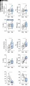
Figure 2. A single dose of Set increases total oxygen consumption (VO2) and carbon dioxide production (VCO2) compared with Veh, but VE increases out of proportion to increases in VO2 and VCO2.
Data in DIO male (top panels, n = 14), lean male (middle panels, n = 9–13), and DIO female mice (bottom panels, n = 10–13) are shown. (A) VO2, VCO2, and respiratory exchange ratio (RER) throughout the light phase. (B) VE/VO2 and VE/VCO2. *P < 0.05, **P < 0.01, ***P < 0.001, ****P < 0.0001 using a paired 2-tailed t test or Wilcoxon’s matched-pairs, signed-rank test.
Figure 3
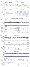
Figure 3. Representative respiratory recordings in DIO male mice treated with a single dose of Set or Veh.
(A) Set increased minute ventilation in room air, increasing RR, and enhanced the HCVR during wakefulness. (B) Obese mice showed inspiratory flow limitation (asterisks) in the presence of increased respiratory effort during sleep, which is a hallmark manifestation of upper airway obstruction. A representative tracing of NREM sleep in a mouse treated with Veh is shown. Outlined fragments on the left are expanded on the right. (C) Several central apneas with oxyhemoglobin desaturations during NREM sleep in an obese mouse treated with Veh (left), which significantly improved after Set treatment (right). EEG, electroencephalogram; EMG, nuchal electromyogram; SpO2, oxyhemoglobin saturation; INSP, inspiration; EXPIR, expiration. On the respiratory tracings, inspirations are down and expirations are up. a.u., arbitrary units.
Figure 4
Figure 4. Set increases ventilation during sleep.
(A and B) Sleep studies showed that Set significantly increased maximal inspiratory flow (VImax), minute ventilation (VE), and respiratory rate (RR) during NREM (A) and REM (B) sleep in male DIO mice. (C and D) Set nearly abolished apneas (C) and improved the oxyhemoglobin desaturation index (ODI; ≥4% from pulse oximetry baseline) (D). VT, tidal volume. n = 7–9. *P ≤ 0.05 using the Wilcoxon’s matched-pairs, signed-rank test.
Figure 5
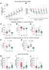
Figure 5. Set treatment for 2 weeks augments the HCVR.
(AE) Set decreased food intake (A), induced mild weight loss (*P < 0.05, Set vs. Veh, P < 0.05, Set vs. pair-fed [PF] using fitted linear mixed models and Tukey’s multiple comparison) (B), increased RR (C) but not VT (D), and increased VE in room air (E). (F and G) Set increased VE at 8% CO2 (F) and HCVR (G) in DIO male C57BL/6J mice in comparison with Veh and PF (*P ≤ 0.05, **P < 0.01, ***P < 0.001, using 1-way ANOVA and Tukey’s multiple-comparison test). (HJ) Set decreased apnea index during NREM (H), REM (I), and total (J) sleep (*P < 0.05 using fitted robust linear models). n = 9 in the Set group; n = 8 in the Veh group; n = 9 in PF group.
Figure 6
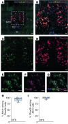
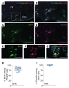
Figure 6. Mc4r is expressed in Phox2b+ neurons of the NTS.
(AG) Lower-power (A) and higher-power (BG) images of the NTS with DAPI, Mc4r, and Phox2b probes and merged images. (H) Percentage of Phox2b+ cells expressing Mc4r in the NTS. (I) Percentage of Mc4r+ cells expressing Phox2b in the NTS. CC, central canal; XII, hypoglossus nucleus. n = 7–8 mice. Scale bars: 50 μm (A); 30 μm (BD); 20 μm (EG).
Figure 7
Figure 7. Mc4r is expressed in the Phox2b+ neurons of the retrotrapezoid nucleus.
(AG) Lower-power (A) and higher-power (BG) images of the retrotrapezoid nucleus (RTN) with DAPI, Mc4r, and Phox2b probes and merged images. (H) Percentage of Phox2b+ cells expressing Mc4r in the RTN. (I) Percentage of Mc4r+ cells expressing Phox2b in the RTN. 7N, facial nucleus. n = 7–8 mice. Scale bars: 50 μm (AD); 15 μm (EG).
Figure 8
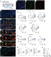
Figure 8. Chemogenetic stimulation of MC4R+ neurons in the RTN (ventral to the facial motor nucleus) increases VE and the HCVR, but not metabolism.
(A) Cre-dependent DREADDs AAV8-hSyn-DIO-hM3D(Gq)-mCherry was deployed in the RTN of Mc4r-Cre DIO mice. (BG) In situ hybridization images. (B) Lower-power image of the brainstem containing RTN. (C) High-power image of the ventral surface of the brainstem containing Mc4r/Nmb/mCherry/DAPI. The outlined area is enlarged in DF. The arrowheads point to Nmb and mCherry (E) and Mc4r and mCherry (F) colocalizations. (G) Histology in the presence of Cre-dependent caspase (AAV5-flex-taCasp3-TEVp) (Casp). Note a significant reduction in Mc4r and Nmb. (HJ) Individual in situ hybridization images showing DAPI in blue (H), Mc4r in green (I), and mCherry in red (J). (KM) Upon stimulation with the DREADDs ligand J60, mice showed increases in VT and VE (K) and HCVR (L) without any effect on oxygen consumption (VO2), CO2 production (VCO2), or the respiratory exchange ratio (RER) (M) in comparison with saline (Sal). n = 5–7. *P ≤ 0.05, using the Wilcoxon’s matched-pairs, signed-rank test. (N) Percentage of colocalized cells. (O) Number of Nmb+ among Mc4r+ cells. **P < 0.01, using the Mann-Whitney U test (n = 5 – 6). 7N, facial motor nucleus. Sp5, spinal trigeminal nucleus. Scale bars: 500 μm (B); 100 μm (C); 50 μm (DF); 30 μm (G); 100 μm (HJ).
Figure 9
Figure 9. Cre-dependent caspase (AAV5-flex-taCasp3-TEVp) (Casp) administered to the RTN of Mc4r-Cre mice abolishes the effect of Set on the HCVR.
Elimination of MC4R+ neurons in the RTN did not affect breathing at normocapnic conditions, and the stimulating effect of Set on RR was still present (A); but the effects of Set on VE at 8% CO2 and HCVR were abolished (B). n = 9. *P ≤ 0.05, **P < 0.01, using the Wilcoxon’s matched-pairs, signed-rank test.
Figure 10
Figure 10. Parafacial MC4R+ neurons are synaptically connected to the rVRG neurons, which innervate phrenic motoneurons.
(A) Cre-dependent ChR2-EYFP was deployed in the parafacial region of Mc4r-Cre mice, and the the C3–C4 segments of the spinal cord were injected with cholera toxin B (CTB) Alexa Fluor 555. (B) Lower-power image shows extensive density of MC4R+ green fibers surrounding CTB+ red cells (top panel). Higher-power images of the same region show (from left to right) MC4R+ fibers, CTB+ neurons, and a merged image showing that green MC4R+ fibers enmesh the red CTB+ neurons (bottom panels). Scale bars: 100 μm (top panel) and 50 μm (bottom panels). (C) Conceptual paradigm describing how MC4R+ neurons stimulate breathing. SC, spinal cord.

References

    1. Mokhlesi B. Obesity hypoventilation syndrome: a state-of-the-art review. Respir Care. 2010;55(10):1347–1362. - PubMed
    1. Nowbar S, et al. Obesity-associated hypoventilation in hospitalized patients: prevalence, effects, and outcome. Am J Med. 2004;116(1):1–7. doi: 10.1016/j.amjmed.2003.08.022. - DOI - PubMed
    1. Gastaut H, et al. Polygraphic study of the episodic diurnal and nocturnal (hypnic and respiratory) manifestations of the Pickwick syndrome. Brain Res. 1966;1(2):167–186. doi: 10.1016/0006-8993(66)90117-X. - DOI - PubMed
    1. Remmers JE, et al. Pathogenesis of upper airway occlusion during sleep. J Appl Physiol Respir Environ Exerc Physiol. 1978;44(6):931–938. - PubMed
    1. Peppard PE, et al. Increased prevalence of sleep-disordered breathing in adults. Am J Epidemiol. 2013;177(9):1006–1014. doi: 10.1093/aje/kws342. - DOI - PMC - PubMed

MeSH terms