. 2025 Feb 25;122(8):e2420383122.
doi: 10.1073/pnas.2420383122. Epub 2025 Feb 18.

A circRNA-mRNA pairing mechanism regulates tumor growth and endocrine therapy resistance in ER-positive breast cancer

Affiliations

A circRNA-mRNA pairing mechanism regulates tumor growth and endocrine therapy resistance in ER-positive breast cancer

Jia Yi et al. Proc Natl Acad Sci U S A. .

Abstract

The molecular mechanisms underlying estrogen receptor (ER)-positive breast carcinogenesis and drug resistance remain incompletely understood. Elevated expression of CCND1 is linked to enhanced invasiveness, poorer prognosis, and resistance to drug therapies in ER-positive breast cancer. In this study, we identify a highly expressed circular RNA (circRNA) derived from FOXK2, called circFOXK2, which plays a key role in stabilizing CCND1 mRNA, thereby promoting cell cycle progression, cell growth, and endocrine therapy resistance in ER-positive breast cancer cells. Mechanistically, circFOXK2 binds directly to CCND1 mRNA via RNA-RNA pairing and recruits the RNA-binding protein ELAVL1/HuR, stabilizing the CCND1 mRNA and enhancing CCND1 protein levels. This results in activation of the CCND1-CDK4/6-p-RB-E2F signaling axis, driving the transcription of downstream E2F target genes and facilitating the G1/S transition during cell cycle progression. Notably, targeting circFOXK2 with antisense oligonucleotide (ASO-circFOXK2) suppresses ER-positive breast cancer cell growth both in vitro and in vivo. Moreover, combination therapy with ASO-circFOXK2 and tamoxifen exhibits synergistic effects and restores tamoxifen sensitivity in tamoxifen-resistant cells. Clinically, high circFOXK2 expression is positively correlated with CCND1 levels in both ER-positive breast cancer cell lines and patient tumor tissues. Overall, our findings reveal the critical role of circFOXK2 in stabilizing the oncogene CCND1 and promoting cancer progression, positioning circFOXK2 as a potential therapeutic target for ER-positive breast cancer in clinical settings.

Keywords: CCND1; RNA-RNA pairing; breast cancer; circFOXK2; endocrine therapy resistance.


Conflict of interest statement

Competing interests statement:The authors declare no competing interest.

Figures

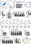
Fig. 1.
Fig. 1.
CircFOXK2 is required for ER-positive breast cancer cell growth both in vitro and in vivo. (A) Standard PCR was performed by using divergent primer sets flanking the junction region of circFOXK2, followed by Sanger sequencing. The sequences flanking the junction region are shown. The junction site is highlighted in light blue. The Sanger sequencing histogram is shown at the bottom. D1: divergent primer 1; D2: divergent primer 2. (BD) MCF7 cells transfected with control siRNA (si-CTL) or two independent siRNAs specifically targeting circFOXK2 (si-circFOXK2-1 and si-circFOXK2-2) were subjected to RT-qPCR (B), cell proliferation (C), and FACS (D) analysis (±SD, **P < 0.01, ***P < 0.001). (EG) MCF7 cells transfected with empty vector (EV) or vector expressing circFOXK2 were subjected to RT-qPCR (E), cell proliferation (F), and FACS (G) analysis (±SD, ***P < 0.001). (H) MCF7 cells infected with control shRNA (sh-CTL) or two independent shRNAs specifically targeting circFOXK2 (sh-circFOXK2-1 and sh-circFOXK2-2) were subjected to RT-qPCR (±SD, ***P < 0.001). (IK) MCF7 cells infected with sh-CTL, sh-circFOXK2-1, or sh-circFOXK2-2 as described in (H) were injected subcutaneously into female BALB/C nude mice for xenograft assay. The growth curve (I), image (J), and weight (K) of tumors are shown (n = 5, ±SD, **P < 0.01, ***P < 0.001).
Fig. 2.
Fig. 2.
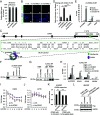
CircFOXK2 positively regulates the expression of CCND1 to activate E2F target genes and promote G1/S transition. (A) MCF7 cells were transfected with control siRNA (si-CTL) or two individual siRNAs specific against circFOXK2 (si-circFOXK2-1 and si-circFOXK2-2) followed by RNA-seq analysis. The correlation between the effects of the two si-circFOXK2 on the whole transcriptome is shown (Pearson correlation coefficient = 0.7717). (B) Genes that are positively and negatively regulated by circFOXK2 based on RNA-seq analysis as described in (A) are shown by volcano plot (FDR < 0.05, FC ≥ 1.5). (C) The five most enriched GO terms for genes positively regulated by circFOXK2 are shown. (D) UCSC genome browser view of RNA-seq as described in (A) for CCND1 is shown. (EG) MCF7 cells transfected with si-CTL, si-circFOXK2-1, or si-circFOXK2-2 were subjected to RT-qPCR (E and G) and immunoblotting (IB) (F) analysis (±SD, *P < 0.05, **P < 0.01, ***P < 0.001). Molecular weight is indicated (Kilodalton: kDa). (H) Tumor samples as described in Fig. 1J were subjected to RNA extraction and RT-qPCR analysis (n = 5, ±SD, *P < 0.05, **P < 0.01, ***P < 0.001). (IK) MCF7 cells transfected with si-CTL or si-circFOXK2 in the presence of EV or vector expressing circFOXK2 were subjected to RT-qPCR (I and K) and IB (J) analysis (±SD, *P < 0.05, **P < 0.01, ***P < 0.001). (LO) MCF7 cells transfected with si-CTL or si-circFOXK2 in the presence of EV or vector expressing Flag-tagged CCND1 were subjected to IB (L), RT-qPCR (M), FACS (N), and cell proliferation (O) analysis (±SD, *P < 0.05, **P < 0.01, ***P < 0.001).
Fig. 3.
Fig. 3.
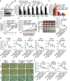
CircFOXK2 pairs with the 3′ UTR of CCND1 mRNA and recruits ELAVL1 to stabilize CCND1 mRNA. (A) MCF7 cells were subjected to cellular fractionation followed by RNA extraction and RT-qPCR analysis for genes as indicated. (B) MCF7 cells transfected with control siRNA (si-CTL) or two individual siRNAs specific against circFOXK2 (si-circFOXK2-1 and si-circFOXK2-2) were subjected to RNA-FISH analysis using probes specifically targeting circFOXK2. Nuclei was stained with DAPI. Green: circFOXK2; Blue: DAPI. (Scale bar, 5 μm.) (C) Schematic representation of the predicted RNA–RNA interaction regions between circFOXK2 and the 3′ UTR of CCND1. (D) In vitro RNA–RNA interaction assay was performed by incubating streptavidin C1 Dynabeads with or without the 3′ UTR of CCND1 in the presence or absence of biotin-labeled, sense circFOXK2 (circFOXK2-S), antisense circFOXK2 (circFOXK2-AS), or sense circFOXK2 with the predicated CCND1-binding region deleted (circFOXK2 (∆120-217)-S). The precipitated RNAs were measured by RT-qPCR analysis using primers specifically targeting the 3′ UTR of CCND1. Data were normalized to the amount of RNA pulled down from empty beads (±SD, **P < 0.01, ***P < 0.001). (E and F) ChIRP assay was performed by incubating cell lysates prepared from MCF7 cells with or without sense (Junction-S) or antisense (Junction-AS) probe specifically targeting the junction region of circFOXK2 followed by RT-qPCR (E) and IB (F) analysis (±SD, n.s: nonsignificant, ***P < 0.001). (G and H) RIP assay was performed by incubating cell lysates prepared from MCF7 cells transfected with si-CTL or si-circFOXK2 with control IgG or anti-ELAVL1 antibody, followed by IB (G) and RT-qPCR (H) analysis (±SD, n.s: nonsignificant, *P < 0.05, **P < 0.01). (I and J) MCF7 cells were transfected with si-CTL or si-circFOXK2 and then treated with or without Actinomycin D (ActD, 10 μg/mL) for duration as indicated, followed by RT-qPCR analysis to examine the expression of CCND1 (I) and ACTIN (J) (±SD, *P < 0.05, **P < 0.01). (K and L) HEK293T cells were transfected with EV or vector expressing circFOXK2 in the presence or absence of Flag-tagged, wild-type (WT) CCND1 or CCND1 with the circFOXK2-binding region in the 3′ UTR deleted (∆2878–2963) for 48 h followed by RT-qPCR (K) and IB (L) analysis (±SD, **P < 0.01, ***P < 0.001).
Fig. 4.
Fig. 4.
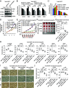
ASO targeting circFOXK2 suppresses ER-positive breast cancer cell growth both in vitro and in vivo. (AE) MCF7 cells transfected with control ASO (ASO-CTL) or ASO specifically targeting circFOXK2 (ASO-circFOXK2) were subjected to RT-qPCR (A and C), IB (B), FACS (D), and cell proliferation (E) analysis (±SD, *P < 0.05, **P < 0.01, ***P < 0.001). (FH) Mice inoculated subcutaneously with MCF7 cells were treated with ASO-CTL or ASO-circFOXK2 (5 nmol per dose, every 3 d) intratumorally for six times. The growth curve (F), image (G), and weight (H) of tumors are shown (n = 5, ±SD, **P < 0.01, ***P < 0.001). (I) Tumor samples as described in (G) were subjected to RNA extraction and RT-qPCR analysis (n = 5, ±SD, *P < 0.05, **P < 0.01, ***P < 0.001).
Fig. 5.
Fig. 5.
Combination treatment with ASO-circFOXK2 and tamoxifen shows synergistic effects in suppressing ER-positive breast cancer cell growth both in vitro and in vivo. (AC) MCF7 cells were transfected with control ASO (ASO-CTL) or ASO specifically targeting circFOXK2 (ASO-circFOXK2) for 36 h before treating with or without tamoxifen (Tam, 5 μM) for 36 h, followed by IB (A), RT-qPCR (B), and FACS (C) analysis (±SD, *P < 0.05, **P < 0.01, ***P < 0.001). (D) MCF7 cells were transfected with ASO-CTL or ASO-circFOXK2 and then treated with or without tamoxifen (Tam, 5 μM), followed by cell proliferation assay (±SD, **P < 0.01, ***P < 0.001). (EG) Female BALB/C nude mice were inoculated subcutaneously with MCF7 cells. Once the tumors were palpable, mice were treated intratumorally with either ASO-CTL or ASO-circFOXK2 (2.5 nmol per dose, every 3 d for six cycles). Tamoxifen (Tam, 20 mg/kg, every 3 d for six cycles) or vehicle were administrated intragastrically (i.g.) every other day. The growth curve (E), image (F), and weight (G) of tumors are shown (n = 5, ±SD, **P < 0.01, ***P < 0.001). (H) Tumor samples as described in (F) were subjected to RNA extraction and RT-qPCR analysis (n = 5, ±SD, *P < 0.05, **P < 0.01, ***P < 0.001). (I) Tumor samples as described in (F) were subjected to IHC analysis with antibodies against Ki67, CCND1, or p-RB, and representative images are shown (400× magnification). (Scale bar, 50 μm.) (J) The average optical density (OD) of the tumor areas as described in (I) was assessed using ImageJ, and results are presented as the relative staining intensity compared to control (n = 5, ±SD, *P < 0.05, **P < 0.01, ***P < 0.001).
Fig. 6.
Fig. 6.
ASO-circFOXK2 resensitizes tamoxifen-resistant ER-positive breast cancer cells to tamoxifen treatment. (AC) Tamoxifen-resistant MCF7 (TamR-MCF7) cells were transfected with control ASO (ASO-CTL) or ASO specifically targeting circFOXK2 (ASO-circFOXK2) for 36 h before treating with or without tamoxifen (Tam, 5 μM) for 36 h, followed by IB (A), RT-qPCR (B), and FACS (C) analysis (±SD, n.s: nonsignificant, *P < 0.05, **P < 0.01, ***P < 0.001). (D) TamR-MCF7 cells were transfected with ASO-CTL or ASO-circFOXK2 and treated with or without tamoxifen (Tam, 5 μM), followed by cell proliferation assay (±SD, n.s: nonsignificant, *P < 0.05, ***P < 0.001). (EG) Female BALB/C nude mice were inoculated subcutaneously with TamR-MCF7 cells. Once the tumors were palpable, mice were treated intratumorally with either ASO-CTL or ASO-circFOXK2 (2.5 nmol per dose, every 3 d for six cycles). Tamoxifen (Tam, 20 mg/kg, every 3 d for six cycles) or vehicle were administrated intragastrically (i.g.) every other day. The growth curve (E), image (F), and weight (G) of tumors are shown (n = 5, ±SD, n.s: nonsignificant, **P < 0.01, ***P < 0.001). (H) Tumor samples as described in (F) were subjected to RNA extraction and RT-qPCR analysis (n = 5, ±SD, n.s: nonsignificant, *P < 0.05, **P < 0.01, ***P < 0.001). (I) Tumor samples as described in (F) were subjected to IHC analysis with antibodies against Ki67, CCND1, or p-RB, and representative images are shown (400× magnification). (Scale bar, 50 μm.) (J) The average OD of the tumor areas as described in (I) was assessed using ImageJ, and results are presented as the relative staining intensity compared to control (n = 5, ±SD, n.s: nonsignificant, *P < 0.05, **P < 0.01, ***P < 0.001).
Fig. 7.
Fig. 7.
CircFOXK2 is highly expressed and positively correlated with CCND1 in ER-positive breast cancer as well as other cancer types. (A) The expression of circFOXK2 was analyzed by RT-qPCR in a normal breast epithelial cell line and different subtypes of breast cancer cell lines as indicated (±SD, **P < 0.01, ***P < 0.001). (B and C) The expression of circFOXK2 (B) and CCND1 (C) was analyzed by RT-qPCR in paired ER-positive (ER+, n = 26) or ER-negative (ER, n = 15) breast tumor and adjacent normal breast tissues (±SD, *P < 0.05, **P < 0.01). (DF) The correlation between the expression of circFOXK2 and CCND1 in breast cancer cell lines (D) as well as ER+ (E) and ER (F) breast tumor tissues is shown. (G and H) The expression of circFOXK2 (G) and CCND1 (H) was analyzed by RT-qPCR in paired CRC (n = 14), ESCC (n = 14), and PRAD (n = 14) tumor and adjacent normal tissues (±SD, *P < 0.05, **P < 0.01). (I) The correlation between the expression of circFOXK2 and CCND1 in CRC, ESCC, and PRAD tumor tissues is shown. (J) A proposed model depicts that elevated circFOXK2 in ER-positive breast cancer cells directly binds to the 3′ UTR of CCND1 mRNA and recruits ELAVL1 to stabilize CCND1 mRNA, leading to the accumulation of CCND1 protein, uncontrolled G1/S progression, and eventually breast tumorigenesis and tamoxifen resistance.

Similar articles

Cited by

References

    1. Bray F., et al. , Global cancer statistics 2022: GLOBOCAN estimates of incidence and mortality worldwide for 36 cancers in 185 countries. CA Cancer J. Clin. 74, 229–263 (2024), 10.3322/caac.21834. - DOI - PubMed
    1. Nolan E., Lindeman G. J., Visvader J. E., Deciphering breast cancer: From biology to the clinic. Cell 186, 1708–1728 (2023), 10.1016/j.cell.2023.01.040. - DOI - PubMed
    1. Will M., Liang J., Metcalfe C., Chandarlapaty S., Therapeutic resistance to anti-oestrogen therapy in breast cancer. Nat. Rev. Cancer 23, 673–685 (2023), 10.1038/s41568-023-00604-3. - DOI - PMC - PubMed
    1. Giaquinto A. N., et al. , Breast cancer statistics, 2022. CA Cancer J. Clin. 72, 524–541 (2022), 10.3322/caac.21754. - DOI - PubMed
    1. Ross-Innes C. S., et al. , Differential oestrogen receptor binding is associated with clinical outcome in breast cancer. Nature 481, 389–397 (2012), 10.1038/nature10730. - DOI - PMC - PubMed

MeSH terms