. 2025 Apr 16;16(1):3596.
doi: 10.1038/s41467-025-58361-w.

Structure of human MUTYH and functional profiling of cancer-associated variants reveal an allosteric network between its [4Fe-4S] cluster cofactor and active site required for DNA repair

Affiliations

Structure of human MUTYH and functional profiling of cancer-associated variants reveal an allosteric network between its [4Fe-4S] cluster cofactor and active site required for DNA repair

Carlos H Trasviña-Arenas et al. Nat Commun. .

Abstract

MUTYH is a clinically important DNA glycosylase that thwarts mutations by initiating base-excision repair at 8-oxoguanine (OG):A lesions. The roles for its [4Fe-4S] cofactor in DNA repair remain enigmatic. Functional profiling of cancer-associated variants near the [4Fe-4S] cofactor reveals that most variations abrogate both retention of the cofactor and enzyme activity. Surprisingly, R241Q and N238S retained the metal cluster and bound substrate DNA tightly, but were completely inactive. We determine the crystal structure of human MUTYH bound to a transition state mimic and this shows that Arg241 and Asn238 build an H-bond network connecting the [4Fe-4S] cluster to the catalytic Asp236 that mediates base excision. The structure of the bacterial MutY variant R149Q, along with molecular dynamics simulations of the human enzyme, support a model in which the cofactor functions to position and activate the catalytic Asp. These results suggest that allosteric cross-talk between the DNA binding [4Fe-4S] cofactor and the base excision site of MUTYH regulate its DNA repair function.


Conflict of interest statement

Competing interests: The authors declare no competing interests.

Figures

Fig. 1
Fig. 1. Human MUTYH-TSAC structure and cancer-associated variants within the [4Fe-4S] cluster motif.
A Global structure (shown in ribbons) highlighting functional domains, catalytic residues and the [4Fe-4S] cluster. The simulated annealing composite omit (gray) and anomalous difference (orange) maps were contoured at 1.1- and 6-sigma, respectively. Functional domains and motifs are indicated in different colors. A logo sequence shows the conservation of the cysteine ligands across archaea, bacteria and eukaryote MutY/MUTYH. B Frequency and distribution of pathogenic germline and somatic mutations mapped in MUTYH amino acid sequence. Domains and motifs are indicated with labels below amino acid numbering. Pathogenic somatic mutations (yellow) were obtained from the COSMIC database. Pathogenic germline mutations were acquired from ExAC (blue), gnomAD (red) and TOPMED (green) databases. Reported frequencies were log-normalized (log base 10). Nonsense mutations are indicated with asterisk. Protein-protein interaction sites are shown with black labels. C Structural mapping of cancer-associated variants within the [4Fe-4S] cluster motif.
Fig. 2
Fig. 2. Structural interplay between the [4Fe-4S] cluster and active site.
A Structural composition of the catalytic pocket and proximity to the [4Fe-4S] cluster. The catalytic pocket comprises 4 α-helixes (H1-H4) and a Helix-Helix (H-H) connector. The H-H connector links the α-helix H3 with an α-helix, which is part of both the catalytic pocket, where Asn238 resides, and the [4Fe-4S] cluster. Catalytic Glu134 and Asp236 reside within the α-helix H2 and the H-H connector, respectively. B Mutual evolutionary relationships show a strong coevolutionary signal among the different structural components of the catalytic pocket and the [4Fe-4S] cluster motif of MUTYH/MutY proteins. Mutual Information (MI) detects positions within a multiple sequence alignment that are co-evolving to reveal evolutionary constraints imposed by structure or function. The protein sequence is presented as a circular plot, residue by residue. The different components taken into account by the coevolutionary analysis such as conservation, cumulative mutual information (MI), proximity MI, and MI pairs are depicted in the figure. C Residues implied in the structural connectivity between the [4Fe-4S] cluster and the active site. The H-bond network establishing this connectivity is shown in cyan dotted lines. The simulated annealing composite omit (gray) and anomalous difference (orange) maps were contoured at 1.1- and 6-sigma, respectively. D Disruption of the structural connectivity between the [4Fe-4S] cluster and the active site in the R149Q GsMutY-THF:OG crystal structure. This mutation shows alternative conformations for the [4Fe-4S] cluster with a displacement of 1.4 Å along with two different conformers of Cys 198. R149Q is the analogous R241Q mutation in MUTYH. The simulated annealing composite omit map (gray) was calculated to the 1.51 Å resolution limit and contoured at 1.0-sigma. E Conservation of the residues involved in the [4Fe-4S] cluster-active site structural connectivity among Helix-hairpin-Helix (HhH) DNA glycosylases that also contain the iron-sulfur cofactor (MUTYH/MutY, MIG and EndoIII).
Fig. 3
Fig. 3. Functional impact of [4Fe-4S] cluster cancer-associated variants of MUTYH.
A Total product formation as a function of [MUTYH] from adenine glycosylase assays of cancer-associated [4Fe-4S] cluster variants with OG:A-containing DNA substrates. The OG:A 30-bp duplex (20 nM) was incubated with increasing MBP-MUTYH concentrations (0–250 nM) for 1 h at 37 °C; see “Methods” for assay details B Percent DNA-bound as a function of [MUTYH] determined from electrophoretic mobility shift assays (EMSA) with cancer-associated variants. The THF:OG-containing 30-bp DNA duplex was titrated with increasing concentrations of MBP-MUTYH (0-400 nM absolute concentrations) and incubated for 20 min at 25 °C. Glycosylase and binding experiments were performed in triplicate with error bars representing standard deviation from the average. C Representative storage phosphor autoradiograms of qualitative glycosylase and electrophoretic mobility shift assays (EMSA) with WT, R241Q and N238S MBP-MUTYH mutants. These experiments were carried out as described in Methods. D Mutation frequency measured using rifampicin resistance assay with GT100 muty- mutm- E. coli cells transformed with pMAL-MBP-MUTYH plasmid harboring cancer-associated variations. For each variant, 8 colonies were evaluated in triplicate on rifampicin plates (24 total measurements/variants). The 95% confidence limits based on the mean value are shown in the relevant plots. E CD spectra of purified cancer-associated MBP-MUTYH. Source data are provided as a Source Data file.
Fig. 4
Fig. 4. Kinetics of adenine glycosylase activity and dissociation constants, Kd.
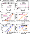
A Full-time course adenine glycosylase assays for determination of kinetic parameters for WT, V246F, and R309C MUTYH. Left panel; time course reaction of MBP-MUTYH adenine excision under single turnover condition ([E] = 100 nM active concentration, [A:OG] = 20 nM). Right panel; kinetics of adenine excision under multiple turnover conditions ([E] = 2.5–5 nM active concentration, [A:OG] = 20 nM). Data fitting was used to obtained the glycosylase (k2) and turnover (k3) rate constants (See “Methods” for more information). B DNA binding isotherms of electrophoretic mobility shift assays (EMSA) for WT, R241Q, V246F and R309C. Left panel; DNA binding isotherms obtained with substrate adenine analog (fA) across OG (10 pM) titrated with increasing concentrations of MBP-MUTYH (0–190 nM). Right panel; DNA binding isotherms obtained with abasic site analog (THF) across OG (10 pM) titrated with increasing concentrations of MBP-MUTYH (0-3000 nM). C DNA binding isotherms obtained for KD determination with fA:OG- and THF:OG-containing DNA duplex binding with WT, R241Q and N238S MBP-MUTYH. Data for R241Q are shown in both panel B and C for comparison. All experiments were repeated in triplicate and the error bars the standard deviation from the mean. Source data are provided as a Source Data file.
Fig. 5
Fig. 5. Molecular dynamics simulation.
Energy decomposition (left), normal mode (middle), and network (right) analyses based on the MD trajectories are shown. The left and right panels show a close-up view of the residues that participate in the structural interplay between the [4Fe-4S] cluster and the active site along with values for energy decomposition analysis (EDA, left) and network analysis results (right). The EDA reports the intermolecular non-bonded interactions (Coulomb and van der Waals interactions; kcal/mol) involving Asn238 or Arg241 (black and red values, respectively). Below, the total energy contribution for the system is shown for Asn238 (black) and Arg241 (red) and corresponding cancer-associated mutants. For the network analysis the betweenness of the nodes involved in the multi-motif bridge is shown in brown (right-upper panel) and the optimal path between the AP site and [4Fe-4S] cluster is displayed in magenta (right-lower panel). The first normal mode is shown in the middle where the MUTYH and DNA are shown in blue and gray, respectively.
Fig. 6
Fig. 6. Summary of functional impacts of MUTYH [4Fe-4S] CAVs.
The different functional impacts of MUTYH cancer-associated variants in proximity to the [4Fe-4S] cluster allowed for grouping into distinct classes.  The impact statements correlate with the color of the residue where the variants are localized.
Fig. 7
Fig. 7. Proposed catalytic mechanism for MUTYH and disrupted mechanism in R241Q and N238S cancer-associated variants.
The adenine excision catalyzed by MUTYH is initiated with the protonation of the adenine by Glu134 which is followed by the glycosidic bond cleavage and formation of an oxacarbenium ion intermediate. Asp236 is proposed to attack the oxacarbenium ion at C1’ to stabilize it by a covalent intermediate. Finally, Glu134 activates a water molecule for nucleophilic attack at C1 to form the AP site product. The structural interplay between the [4Fe-4S] cluster is important for step 2 of catalysis, where Asp236 attacks the oxacarbenium ion. Asp236 has to be oriented near the N-glycosidic bond and deprotonated for the nucleophilic attack. Such orientation and deprotonation status of Asp236 is modulated by the H-bond with Gln238 and allosterically through the [4Fe-4S] cluster via the Cys290-Arg241-Asn238-Asp236 structural bridge. Upon R241Q and N238S mutations the H-bond between Asp236 and Asn238 becomes unstable (cyan dashed arrow) or is broken (black curved lines). With the structural bridge thus compromised the position, protonation state, and charge of catalytic Asp236 position are also impacted (double-headed dashed arrows) with the net consequence of impeding nucleophilic attack of the oxacarbenium ion. This pathological scenario is accompanied by structural instability of the [4Fe-4S] cluster and loss of DNA-MUTYH interactions.

References

    1. Al-Tassan, N. et al. Inherited variants of MYH associated with somatic G: C→ T: a mutations in colorectal tumors. Nat. Genet.30, 227–232 (2002). - PubMed
    1. Raetz, A. G. & David, S. S. When you’re strange: Unusual features of the MUTYH glycosylase and implications in cancer. DNA Repair80, 16–25 (2019). - PMC - PubMed
    1. Engstrom, L. M. et al. A zinc linchpin motif in the MUTYH glycosylase interdomain connector is required for efficient repair of DNA damage. J. Am. Chem. Soc.136, 7829–7832 (2014). - PMC - PubMed
    1. Messick, T. E. et al. Noncysteinyl coordination to the [4Fe-4S] 2+ cluster of the DNA repair adenine glycosylase MutY introduced via site-directed mutagenesis. Structural characterization of an unusual histidinyl-coordinated cluster. Biochemistry41, 3931–3942 (2002). - PubMed
    1. Porello, S. L., Leyes, A. E. & David, S. S. Single-turnover and pre-steady-state kinetics of the reaction of the adenine glycosylase MutY with mismatch-containing DNA substrates. Biochemistry37, 14756–14764 (1998). - PubMed