. 2025 Apr 15;16(1):300.
doi: 10.1038/s41419-025-07611-2.

A variant of the autophagic receptor NDP52 counteracts phospho-TAU accumulation and emerges as a protective factor for Alzheimer's disease

Affiliations

A variant of the autophagic receptor NDP52 counteracts phospho-TAU accumulation and emerges as a protective factor for Alzheimer's disease

Anna Mattioni et al. Cell Death Dis. .

Abstract

Selective elimination of early pathological TAU species may be a promising therapeutic strategy to reduce the accumulation of TAU, which contributes to neurodegeneration and is a hallmark of Alzheimer's disease (AD). Pathological hyper-phosphorylated TAU can be degraded through selective autophagy, and NDP52/CALCOCO2 is one of the autophagy receptors involved in this process. In 2021, we discovered a variant of NDP52, called NDP52GE (rs550510), that is more efficient at promoting autophagy. We here anticipate that this variant could be a powerful factor that could eliminate pathological forms of TAU better than its WT form (NDP52WT). Indeed, we provide evidence that in in vitro systems and in a Drosophila melanogaster model of TAU-induced AD, the NDP52GE variant is much more effective than the NDP52WT in reducing the accumulation of pathological forms of TAU through the autophagic process and rescues typical neurodegenerative phenotypes induced by hTAU toxicity. Mechanistically, we showed that NDP52WT and NDP52GE bind pTAU with comparable efficiency, but that NDP52GE binds the autophagic machinery (LC3C and LC3B) more efficiently than NDP52WT does, which could explain its greater efficiency in removing pTAU. Finally, by performing a genetic analysis of a cohort of 435 AD patients, we defined the NDP52GE variant as a protective factor for AD. Overall, our work highlights the variant NDP52GE as a resilience factor in AD that shows a robust effectiveness in driving pathological TAU degradation.


Conflict of interest statement

Competing interests: The authors declare no competing interests. Ethics approval and consent to participate: The patients from IRCCS Santa Lucia Foundation Hospital of Rome were enrolled for a previous study approved by the Ethical Committee (CE/PROG.650 approved on 01/03/2018). The patients from the memory clinic of CRIC in Padua were collected according to the study protocol “Analisi delle Basi Biologiche della Malattia di Alzheimer eredofamiliare e delle forme sporadiche tardive” (3950/AO/2016) approved by the local ethic committee of the recruitment center on October 06th, 2016. All peripheral blood samples were collected after obtaining the written informed consent of the study participants, in accordance with the Helsinki Declaration.

Figures
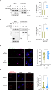
Fig. 1
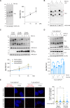
Fig. 1. NDP52GE binds LC3C more efficiently than NDP52WT in a human neuroblastoma cell line.
A Lysates of SH-SY5Y cells expressing the indicated FLAG- and HA-tagged proteins was immunoprecipitated with anti-HA beads. Samples were analyzed by Western blot using the indicated antibodies. The graph reports the amount of the indicated FLAG-NDP52 protein coprecipitated by the corresponding HA-LC3C protein. Data were expressed as percentage variation over FLAG-NDP52WT. Images and data are representative of seven independent experiments. B Lysates of SH-SY5Y cells expressing the indicated GFP- and HA-tagged proteins were immunoprecipitated with anti-GFP beads. Samples were analyzed by Western blot using the indicated antibodies. The graph reports the amount of HA-LC3C coprecipitated by the corresponding GFP-NDP52 protein. Data were expressed as percentage variation over GFP-NDP52WT. Images and data are representative of four independent experiments. C Representative confocal images of SH-SY5Y cells expressing the indicated GFP- and HA-tagged proteins, fixed and stained with anti-HA (magenta staining) antibodies and with DAPI (blue staining in the merge panels) to detect nuclei. Colocalization of GFP-NDP52 and HA-LC3C was quantified by measuring the Pearson’s Correlation Coefficient. The results are reported in the graph. Images are representative of three independent experiments, and at least 30 cells for each condition were analyzed. Scale bar: 10 μm. In (B, C) data are presented as means ± SEM. *p < 0,05; ***p = <0.001 (two-tailed unpaired t-test).
Fig. 2
Fig. 2. The increased binding affinity of NDP52GE towards LC3C strictly depends on the cLIR motif.
A Schematic representation of the human NDP52WT protein, the human variant NDP52GE, and the NDP52 mutants used in this study. The G140E aminoacidic substitution is highlighted, and the position relative to cLIR motif as well as the LIR-like motif are shown in the expanded dotted area. SKICH skeletal muscle and kidney-enriched inositol phosphatase carboxyl homology, cLIR non-canonical LC3-interacting region, LIR LC3-interacting region, UBZ ubiquitin-binding zinc finger. Lysates of SH-SY5Y cells expressing the indicated FLAG- and HA-tagged proteins were immunoprecipitated with anti-HA (B) or anti-FLAG (C) beads. Samples were analyzed by Western blot using the indicated antibodies. The graphs report the amount of the indicated FLAG-NDP52 protein coprecipitated by the corresponding HA-LC3C protein. Data were expressed as percentage variation over FLAG-NDP52WT. Images and data are representative of three independent experiments. ****p-value < 0.0001; ***p-value < 0.001; **p-value < 0.01: *p-value < 0.05 (Ordinary One-way ANOVA, Turkey’s multiple comparisons test).
Fig. 3
Fig. 3. NDP52GE reduces pTAU levels better than NDP52WT, in a human neuroblastoma cell line.
A Western blot (WB) on extracts of SH-SY5Y cells treated with 100 nM OkA for the indicated times. TAU bands were measured, summed, and normalized to the corresponding signal of VINCULIN. Results are expressed as percentage variation of the levels at time 0. Images and data are representative of eight independent experiments. B Lysates of SH-SY5Y cells treated as in (A) were split and incubated with (+) or without (−) Lambda Protein Phosphatase (λPP). Images are representative of three independent experiments. Lilac lines = non-phosphorylated TAU; purple lines = phosphorylated TAU. C SH-SY5Y cells expressing the indicated FLAG-tagged proteins were treated as in (A). Signals from TAU antibody were measured on WB and normalized as in (A). Results are expressed as percentage variation of the levels measured at time 0 for each FLAG-tagged protein expressing group. Images and data are representative of eight independent experiments and are reported as means ± SEM. * indicates comparison to time 0 of each FLAG-tagged protein expressing group; & indicates comparison between 1 h and 2 h of each group (two tailed unpaired t-test) D SH-SY5Y cells expressing FLAG-tagged NDP52GE were pre-treated with NH4Cl (20 mM, 24 h) or not, and then were treated with 100 nM OkA as indicated. Signals from TAU antibody were measured on WB and normalized as in (A). Images and data are representative of three independent experiments and are reported as means ± SEM. At each OkA time, NH4Cl non-treated vs NH4Cl treated results were tested for statistical significance (two-tailed unpaired t-test). E Representative confocal images of in situ PLA performed in SH-SY5Y overexpressing FLAG-NDP52WT or FLAG-NDP52GE treated with DMSO or with 100 nM OkA for 1.5 h at 37 °C. Dots were counted using CellProfiler software and results were shown in the violin plot graphs. At least 100 cells for each condition were analyzed. (Ordinary One-way ANOVA, Turkey’s multiple comparisons test). Scale bar: 10 μm.
Fig. 4
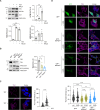
Fig. 4. NDP52GE degrades P301S-TAU via autophagy and is more efficient than NDP52WT.
A WB analysis of human SH-SY5Y P301S-TAU inducible cells non-treated (TET −) or treated (TET +) with tetracycline for 24 h to induce P301S-TAU expression. To block the autophagosome–lysosome fusion, cells were treated with NH4Cl (20 mM, 24 h). Images are representative of three independent experiments. Signals from LC3II, TAU, and NDP52 antibodies were measured and normalized to the corresponding signal of VINCULIN. Data are reported as means ± SEM. (two-tailed unpaired t-test). B WB analysis of human SH-SY5Y P301S-TAU inducible cells overexpressing GFP, GFP-NDP52WT or GFP-NDP52GE and treated with tetracycline to induce P301S-TAU expression. After 16 h, the medium was substituted with a TET-free medium for 8 h in order to evaluate and compare TAU degradation rate. Data are reported as means ± SEM. (two-tailed unpaired t-test). C Representative confocal images of human SH-SY5Y P301S-TAU inducible cells non-treated (TET −) or treated (TET +) with tetracycline for 24 h to induce P301S-TAU expression, fixed and stained with anti-TAU (magenta staining) antibody and with DAPI (blue staining in the merge panels) to detect nuclei. Intensity of anti-TAU signal was measured, and results are reported in the graph. Images are representative of three independent experiments and more than 70 cells for each condition were analyzed. (two-tailed unpaired t-test). Scale bar: 10 μm. D Representative confocal images of human SH-SY5Y P301S-TAU inducible cells overexpressing GFP-NDP52WT or GFP-NDP52GE and treated as in (B). Cells were left untreated (ctrl) or were treated with the autophagic inhibitor 3-Methyladenine (+3-MA) for 8 h. Cells were fixed and stained with anti-TAU antibody and with DAPI to detect nuclei. Intensity of anti-TAU signal was measured, and results are reported in the graph. Images are representative of three independent experiments and more than 200 cells for each condition were analyzed. (two-tailed unpaired t-test).
Fig. 5
Fig. 5. hNDP52GE improves hTAU phenotypes in Drosophila melanogaster better than hNDP52WT.
A Lysates from heads of flies expressing the indicated transgenes in the eye, at 29 °C, under control of GMR-GAL4 driver were analyzed by WB using the indicated antibodies. + refers to non-transgenic flies used as control. Images are representative of four independent experiments. The ratio between dAtg8II/dAtg8I was calculated and reported in the graph as means ± SEM. * indicates comparison of dAtg8II/dAtg8I between flies expressing hNDP52GE and control; § indicates comparison of dAtg8II/dAtg8I between flies expressing hNDP52GE and hNDP52WT (Ordinary One-way ANOVA, Turkey’s multiple comparisons test). B Representative images of eye from flies expressing the indicated human (h) transgenes at 25 °C under control of eyeless-GAL4 driver. Violin plot below reports eye areas. Median and quartiles are shown as dotted lines. More than 150 eyes from both males and females flies were measured for each condition. * indicates comparison to control (+); # indicates comparison to hTAU; § indicates comparison to hTAU;hNDP52WT (Ordinary One-way ANOVA, Turkey’s multiple comparisons test). C Box and whiskers (min to max) plot showing the percentage of flies reaching the threshold. Female flies expressed panneuronally (elav-GAL4) the indicated human transgenes at 29 °C. More than 40 flies per genotype were analyzed (Control n = 54 flies; hTAU n = 44; hTAU, NDP52WT n = 53; hTAU, NDP52GE n = 48 flies). * indicates comparison to control (+); # indicates comparison to hTAU (Ordinary One-way ANOVA, Turkey’s multiple comparisons test). D Survival curve showing lifespan of female flies expressing panneuronally (elav-GAL4) the indicated transgenes at 29 °C. * indicates comparison to control (+); # indicates comparison to hTAU (Log-rank (Mantel-Cox) test). E Lysates from heads of flies expressing panneuronally (elav-GAL4) the indicated transgenes at 29 °C were analyzed by WB using the indicated antibodies. Images are representative of four independent experiments. Signals from the indicated TAU antibodies were measured and normalized to the corresponding signal of GIOTTO. Results are expressed as means ± SEM. # indicates comparison to hTAU; § indicates comparison to hTAU;hNDP52WT (Ordinary One-way ANOVA, Turkey’s multiple comparisons test).
Fig. 6
Fig. 6. NDP52GE is a protective factor in AD and counteracts pTAU accumulation.
Illustration of the highlights of the work. By using two in vitro systems, we showed that the NDP52GE variant counteracts the accumulation of pathological pTAU better than NDP52WT, through the autophagy process. Furthermore, we showed in vivo, in a Drosophila melanogaster model of TAU-mediated AD that the NDP52GE variant partially rescues different pathological phenotypes induced by hTAU expression. Finally, using a genetic association case-control analysis, we showed that NDP52GE is a protective factor in AD. Created with BioRender.com.

Similar articles

Cited by

References

    1. 2022 Alzheimer’s disease facts and figures. Alzheimer’s Dement. 2022;18:700–89. - PubMed
    1. Nichols E, Steinmetz JD, Vollset SE, Fukutaki K, Chalek J, Abd-Allah F, et al. Estimation of the global prevalence of dementia in 2019 and forecasted prevalence in 2050: an analysis for the Global Burden of Disease Study 2019. Lancet Public Health. 2022;7:e105–25. - PMC - PubMed
    1. Madnani RS. Alzheimer’s disease: a mini-review for the clinician. Front Neurol. 2023;14:1–7. - PMC - PubMed
    1. Congdon EE, Ji C, Tetlow AM, Jiang Y, Sigurdsson EM. Tau-targeting therapies for Alzheimer's disease: current status and future directions. Nat Rev Neurol. 2023;19:715–36. - PMC - PubMed
    1. Vogt ACS, Jennings GT, Mohsen MO, Vogel M, Bachmann MF. Alzheimer’s disease: a brief history of immunotherapies targeting amyloid β. Int J Mol Sci. 2023;24:3895. - PMC - PubMed

LinkOut - more resources