. 2025 Apr 15;20(1):50.
doi: 10.1186/s13020-025-01098-x.

Huang-Lian-Jie-Du decoction alleviates cognitive deficits in Alzheimer's disease model 5xFAD mice by inhibiting Trem2/Dap12 signaling pathway

Affiliations

Huang-Lian-Jie-Du decoction alleviates cognitive deficits in Alzheimer's disease model 5xFAD mice by inhibiting Trem2/Dap12 signaling pathway

Rui-Kang Pang et al. Chin Med. .

Abstract

Background: Alzheimer's disease (AD) is a progressive neurodegenerative disorder predominantly affecting the elderly population. It is characterized by cognitive deficits associated with the accumulation of amyloid-beta plaques and neurofibrillary tangles. Huang-Lian-Jie-Du (HLJD) decoction, recognized as a representative formulation with heat-clearing and detoxification effects, has been demonstrated to be effective in treating AD. However, the underlying mechanisms require further investigation.

Methods: 5xFAD mice were administrated low and high doses of HLJD. The Morris water maze test was conducted to assess the effects of HLJD. Aβ42 and total tau protein levels were evaluated. Additionally, network pharmacology analysis was performed to identify therapeutic targets of HLJD's active components and their relevance to AD. ELISA, qPCR, Western Blot, and immunofluorescence assays were employed to confirm the identified pathways. Finally, primary microglia isolated from 5xFAD mice were used to validate the candidate targets of HLJD.

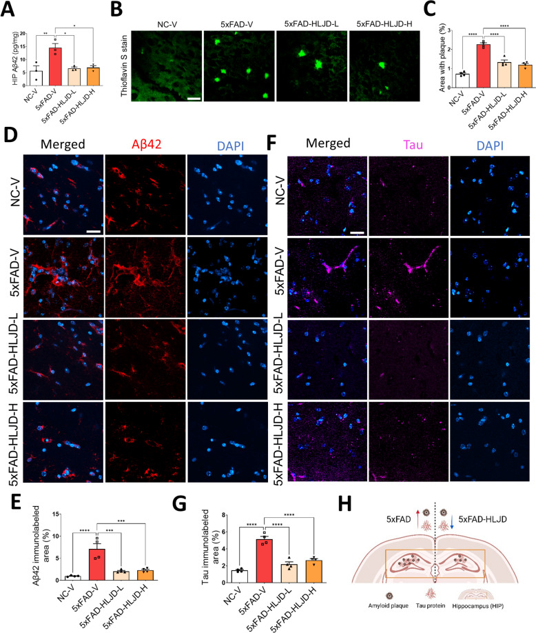
Results: HLJD improved cognitive deficits in 5xFAD mice and reduced amyloid plaque deposition and tau protein levels. Network pharmacology analysis indicated that HLJD influences the neuroinflammatory response, particularly through the Dap12 signaling pathway. This was confirmed by reduced levels of neuroinflammation markers, including TNF-α, IL-1β, IL-6, and indicators of microglial activation and polarization. The expression of Trem2 and Dap12 in the hippocampus (HIP) of 5xFAD mice, as well as in the isolated primary microglia, were downregulated following HLJD treatment.

Conclusion: Our study indicates that HLJD alleviates cognitive deficits in AD by suppressing the Trem2/Dap12 signaling pathway in the HIP of 5xFAD mice, thereby inhibiting microglial neuroinflammation.

Keywords: Alzheimer’s disease; Dap12; Huang-Lian-Jie-Du decoction; Microglia; Neuroinflammation; Trem2.


Conflict of interest statement

Declarations. Ethics approval and consent to participate: The experiment was performed in accordance with the Centralized Animal Facilities of the Hong Kong Polytechnic University Shenzhen Research Institute, with the license number 21-22/122-ABCT-R-OTHERS. Consent for publication: Not applicable. Competing interests: The authors declare that they have no known competing financial interests or personal relationships that could have appeared to influence the work reported in this paper.

Figures

Fig. 1
Fig. 1
HLJD decoction alleviated cognitive impairment in 5xFAD mice. A Experimental timeline. B The escape latencies for both the visible platform on day one and the hidden platform from day 2 to day 5. C Representative behavioral path. D The average latency to target quadrant zone on day 6. E The latency to platform zone on day 6. F The time in target quadrant on day 6. Data are presented as mean ± SEM. *P < 0.05, **P < 0.01, ***P < 0.001, ****P < 0.0001
Fig. 2
Fig. 2
HLJD decoction reduced the expression of Aβ42 and total tau in the HIP of 5xFAD mice. A ELISA analyses for Aβ42 levels in the HIP of mice (n = 3). B Representative images of ThS staining. Scale bars, 25 µm. C Quantification of B (n = 4). D Representative images of Aβ42 staining in the HIP. Scale bars, 25 µm. E Quantification of Aβ42 positive area (n = 4). F Representative images of tau in the HIP. Scale bars, 25 µm. G Quantification of tau positive area (n = 4). H Diagram of the Aβ plaques and total tau protein levels in the HIP of 5xFAD mice treated with or without HLJD decoction. Data are presented as mean ± SEM. *P < 0.05, **P < 0.01, ***P < 0.001, ****P < 0.0001
Fig. 3
Fig. 3
Network pharmacology diagram of HLJD for the treatment of AD. A Venn diagram showing targets of HLJD for AD treatment. B Protein–protein interaction enrichment analysis for the overlapping 230 genes using Metascape. C Network diagram of Traditional Chinese Medicine (yellow color)—active ingredients (green color)—intersecting genes (light yellow color) – disease (red color)
Fig. 4
Fig. 4
Functional enrichment analysis of the interesting target genes. Interesting target genes enriched in the top one cluster from PPI were further analyzed. A GO functional enrichment analysis of the interesting target genes of HLJD in the treatment of AD. B KEGG pathway enrichment analysis of the interesting target genes. C Reactome pathway enrichment analysis of the interesting target genes
Fig. 5
Fig. 5
HLJD decoction ameliorated neuroinflammation in the HIP of 5xFAD mice. AC The mRNA expressions of Tnf-α, Il-1β and Il-6 (n = 3). DF ELISA assays of TNF-α, IL-1β and IL-6 (n = 3). G Representative images of TNF-α. Scale bars, 25 µm. H Quantification of TNF-α immunolabeled area (n = 4). I Representative images of IL-1β. Scale bars, 25 µm. J Quantification of IL1β immunolabeled area (n = 4). K Diagram of the TNF-α, IL-1β and IL-6 levels in the HIP of 5xFAD mice treated with or without HLJD decoction. Data are presented as mean ± SEM. *P < 0.05, **P < 0.01, ***P < 0.001, ****P < 0.0001
Fig. 6
Fig. 6
HLJD decoction alleviated the microglia activation. A Representative images of Iba1 in the HIP. Scale bars, 25 µm. B Microglia coverage (MG) for the Iba1 labelled images within the entire field of view (FOV). C MG volume within the FOV. D Total process length within the FOV. E Number of branch points of Iba1-labeled microglia within the FOV. F Representative images of Iba1 and CD169 staining in the HIP. Scale bars, 25 µm. G Quantification of CD169 immunolabeled area in the HIP. H The percentage of the CD169+ Iba1+ cells in Iba1+ cells (n = 4). Data are presented as mean ± SEM. *P < 0.05, **P < 0.01, ***P < 0.001, ****P < 0.0001
Fig. 7
Fig. 7
HLJD decoction affected microglial polarization and the Trem2/Dap12 signaling pathway. AD The mRNA levels of Il-12a, Nos2, Cxcl10, and Ccl3 in the HIP. EK The mRNA expressions of CD206, Arg-1, Vegf, Il-10, Tgf-β1, Il-4 and Il-13 in the HIP. LO The mRNA expressions of Trem2, Dap12, Csf1 and Csf1r in the HIP (n = 3). Data are presented as mean ± SEM. *P < 0.05, **P < 0.01
Fig. 8
Fig. 8
HLJD decoction inhibited the Trem2/Dap12 signaling pathway in the HIP of 5xFAD mice. A Representative blots of Trem2, Dap12 and β-Actin. B, C Quantification of Trem2 and Dap12 (n = 3). D Representative images of Iba1 and Trem2 staining in the HIP. Scale bars, 25 µm. E Quantification of Trem2 immunolabeled area (n = 4). F Percentage of Trem2+Iba1+ cells in Iba1+ cells (n = 4). G Representative images of Iba1 and Dap12 staining in the HIP. Scale bars, 25 µm. H Quantification of Dap12 immunolabeled area (n = 4). I Percentage of Dap12+Iba1+ cells in Iba1+ cells (n = 4). Data are presented as mean ± SEM. *P < 0.05, ****P < 0.0001
Fig. 9
Fig. 9
HLJD decoction inhibited the Trem2/Dap12 signaling pathway in the primary microglia of 5xFAD mice. A Representative images of IL-1β in the primary microglia of 5xFAD mice. Scale bars, 25 µm. B Quantification of IL-1β immunolabeled area. C Representative images of Trem2 and Iba1 staining in the primary microglia of 5xFAD mice. Scale bars, 25 µm. D Percentage of Trem2+Iba1+ cells in Iba1+ cells. E Representative images of Dap12 and Iba1 staining in the primary microglia of 5xFAD mice. Scale bars, 25 µm. F Percentage of Dap12+Iba1+ cells in Iba1+ cells (n = 3). Data are presented as mean ± SEM. **P < 0.01

References

    1. Ren D, Fu Y, Wang L, Liu J, Zhong X, Yuan J, et al. Tetrandrine ameliorated Alzheimer’s disease through suppressing microg lial inflammatory activation and neurotoxicity in the 5XFAD mouse. Phytomedicine. 2021;90: 153627. - PubMed
    1. Garg N, Choudhry MS, Bodade RM. A review on Alzheimer’s disease classification from normal controls an d mild cognitive impairment using structural MR images. J Neurosci Methods. 2023;384: 109745. - PubMed
    1. Scheltens P, De Strooper B, Kivipelto M, Holstege H, Chételat G, Teunissen CE, et al. Alzheimer’s disease. Lancet. 2021;397:1577–90. - PMC - PubMed
    1. Collaborators GBDDF. Estimation of the global prevalence of dementia in 2019 and forecasted prevalence in 2050: an analysis for the Global Burden of Disease Stud y 2019. Lancet Public Health. 2019;7:e105-e25. - PMC - PubMed
    1. Passeri E, Elkhoury K, Morsink M, Broersen K, Linder M, Tamayol A, et al. Alzheimer’s disease: treatment strategies and their limitations. Int J Mol Sci. 2022;23:13954. - PMC - PubMed

LinkOut - more resources