

VOPP1::EGFR fusion is associated with NFκB pathway activation in a glioneural tumor with histological features of ganglioglioma
- PMID: 40234994
- PMCID: PMC12001695
- DOI: 10.1186/s40478-025-01994-1
VOPP1::EGFR fusion is associated with NFκB pathway activation in a glioneural tumor with histological features of ganglioglioma
Abstract
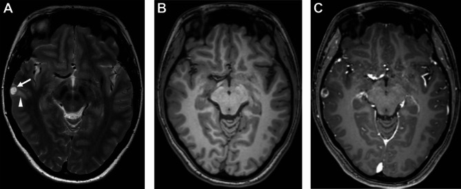
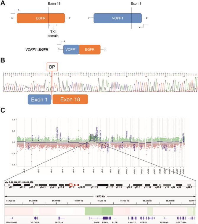
Glioneural tumors are primary brain tumors that consist of both neural and glial neoplastic cells, often presenting with seizures and primarily affecting children and young adults. Specifically, gangliogliomas are composed of neoplastic ganglion and glial cells, accompanied by other characteristic histological features such as lymphoid cuffing, eosinophilic granular bodies, and Rosenthal fibers. Oncogenic driver mutations and gene fusions have been shown to be of prognostic significance in gangliogliomas and can offer potential therapeutic targets. Typical molecular alterations are mitogen-activated protein kinase (MAPK) pathway activations with BRAF p.V600E being the most frequent one. Here, we report for the first time a gene fusion between epidermal growth factor receptor (EGFR) and vesicular, overexpressed in cancer, prosurvival protein 1 (VOPP1) as a potential oncogenic driver in a glioneuronal tumor morphologically resembling ganglioglioma. VOPP1::EGFR fusion associated with the activation of nuclear factor kappa-light-chain-enhancer of activated B cells (NFκB) signaling. Furthermore, we provide histological and epigenetic findings and clinical outcome. The case expands the known molecular spectrum of oncogenic drivers in glioneuronal tumors and provides a link to potentially prognostic and therapeutically relevant alterations.
Keywords: EGFR; Ganglioglioma; Gene fusion; NFκB; VOPP1.
© 2025. The Author(s).
Conflict of interest statement
Declarations. Ethics approval and consent to participate: All procedures were performed in accordance with the ethical standards of the institutional and/or national research committee and with the 1964 Helsinki declaration and its later amendments or comparable ethical standards. The report is based on surgically removed tissue in the context of routine diagnostic workup and represent a retrospective observational study of a single case. Therefore, the Ethics Committee of the medical faculty of the university medical center Leipzig (UKL) has confirmed that no ethical approval is required as regulated by paragraph 15 of the professional regulation of the medical association of Saxony, Germany (§ 15 BO, SLÄK). Consent for scientific usage of tumor tissue and publishing of respective data was obtained from the patient. Consent for publication: The patient has given explicit written consent for scientific usage and publication of clinical, radiological and neuropathological data. Competing interests: The authors declare no competing interests.
Figures


References
-
- Blumcke I, Wiestler OD (2002) Gangliogliomas: an intriguing tumor entity associated with focal epilepsies. J Neuropathol Exp Neurol 61:575–584. 10.1093/jnen/61.7.575 - PubMed
-
- Blumcke I, Spreafico R, Haaker G, Coras R, Kobow K, Bien CG, Pfäfflin M et al (2017) Histopathological findings in brain tissue obtained during epilepsy surgery. N Engl J Med 377:1648–1656. 10.1056/NEJMoa1703784 - PubMed
-
- WHO Classification of Tumours Editorial Board (2021) Central nervous system tumours. https://tumourclassification.iarc.who.int/chapters/45. Accessed 15 Jan 2025
Publication types
MeSH terms
Substances
LinkOut - more resources
Full Text Sources
Medical
Research Materials
Miscellaneous
