Review
. 2025 Dec;19(1):2490308.
doi: 10.1080/19336950.2025.2490308. Epub 2025 Apr 16.

The GluN3-containing NMDA receptors

Affiliations
Review

The GluN3-containing NMDA receptors

Kunlong Xiong et al. Channels (Austin). 2025 Dec.

Abstract

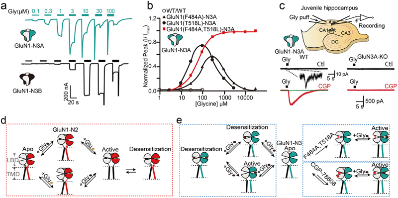
N-methyl-D-aspartate receptors (NMDARs) are heterotetrameric ion channels that play crucial roles in brain function. Among all the NMDAR subtypes, GluN1-N3 receptors exhibit unique agonist binding and gating properties. Unlike "conventional" GluN1-N2 receptors, which require both glycine and glutamate for activation, GluN1-N3 receptors are activated solely by glycine. Furthermore, GluN1-N3 receptors display faster desensitization, reduced Ca2+ permeability, and lower sensitivity to Mg2+ blockage compared to GluN1-N2 receptors. Due to these characteristics, GluN1-N3 receptors are thought to play critical roles in eliminating redundant synapses and pruning spines in early stages of brain development. Recent studies have advanced pharmacological tools for specifically targeting GluN1-N3 receptors and provided direct evidence of these glycine-activated excitatory receptors in native brain tissue. The structural basis of GluN1-N3 receptors has also been elucidated through cryo-EM and artificial intelligence. These findings highlight that GluN1-N3 receptors are not only involved in essential brain functions but also present potential targets for drug development.

Keywords: Ionotropic glutamate receptors; ligand-gated ion channels; pathology and physiology; protein prediction; synaptic transmission.


Conflict of interest statement

No potential conflict of interest was reported by the author(s).

Figures

illustrates the subunit homology of iGluRs, including sequence alignment of GluN1 and GluN3, and schematic models of di- or tri-heteromeric GluN3-containing NMDARs. Panel (a) shows the phylogenetic relationship between iGluR subunits (NMDARs, AMPARs, KARs, and GluDRs) and sequence alignment of GluN1 and GluN3. Panel (b) depicts the schematic structure of GluN1, GluN3A, and GluN3B subunits. Panel (c) presents models of di- and tri-heterotetrameric GluN3-containing NMDARs with “1-3-1-3” or “1-2-1-3” stoichiometries.
Figure 1.
Subunit homology of iGluRs, sequence alignment of GluN1 and GluN3, schematic model of di- or tri-heteromeric GluN3-containing NMDARs. a. The relationship between the iGluRs subunits. The iGluRs are classified into the NMDARs, AMPARs (α-amino-3-hydroxy-5-methyl-4-isoxazole propionic acid receptors), KARs (Kainate receptors), and GluDRs. The sequences Q05586, Q12879.1, Q13224.3, Q14957.3, Q15399.2, Q9R1M7.1, O60391.2, P42261.2, P42262.3, P42263.2, P48058.2, P39086.1, Q13002.1, Q13003.3, Q16099.2, Q16478.2, Q9ULK0.2, O43424.2, were analyzed. b. Schematic structure of the GluN1, GluN3A, and GluN3B subunits. c. Schematic models of several di- and tri-heterotetrameric GluN3-containing NMDARs with “1-3-1-3” or “1-2-1-3” stoichiometries.
demonstrates the spatial and subcellular expression patterns of GluN3. Panel (a) shows a schematic diagram of GluN3 and other NMDAR subunit expressions in the adult brain. Panel (b) displays a schematic representation of changes in GluN3A and GluN3B expression during brain development and comparison of expression in major neuronal cells. Panel (c) includes immunofluorescence and electron micrograph images showing GluN3B expression in cultured neurons, co-localized with synaptophysin, and localized in dendritic spines. Panel (d) illustrates the presynaptic and postsynaptic localization of GluN1-GluN3. Panel (e) presents t-SNE plots showing global expression patterns of Grin1, Grin3a, and Grin3b in the frontal cortex, hippocampus, and thalamus.
Figure 2.
The spatial and subcellular expression pattern of GluN3. a. A schematic diagram illustrates GluN3 and other NMDAR subunits expression pattern in the adult brain. b. Schematic representation of changes in GluN3A and GluN3B expression during brain development and comparison of expression in major neuronal cells. c. GluN3B is expressed in the cultured neurons and co-located with the synaptophysin. Electron micrograph demonstrating GluN3B localized in the dendritic spine. rA/C, recurrent associational-commissural terminal; sp, dendritic spine. d. GluN1-GluN3 can be in both the presynaptic and postsynaptic areas. e. t-distributed stochastic neighbor embedding (t-sne) plots for the frontal cortex, the hippocampus, and the thalamus global expression patterns of Grin1, Grin3a, and Grin3b. Excerpt from [20] (a), [23, 28] (c), [25] (e), and these figures have been reproduced with permission from Oxford University Press (a), Springer nature (c), Elsevier (c), and Frontiers (e), respectively.
explores the structural basis, sequence alignment, and ligand interactions of GluN1-N3 NMDA receptors. Panel (a) shows the structural model of GluN1-GluN3. Panel (b) displays sequence alignment of GluN1, GluN2, and GluN3 subunits, highlighting predicted loops. Panel (c) illustrates ligand-protein interactions of glycine/CGP-78608 with GluN1-LBD and glycine with GluN3A-LBD. Panels (d) and (e) present structural models of glycine-bound and CNQX-bound GluN1-N3A, respectively. Panel (f) shows the density map and structural model of GluN1-N2A-N3A. Panel (g) displays predicted structural models of GluN1-N3A, GluN1-N3B, and GluN1-N3A-N3B.
Figure 3.
Structural basis, sequence alignment, and Ligplot+ analysis of GluN1-N3 NMDA receptors. a. Structural model of GluN1-GluN3. b. Particle sequence alignment of GluN1, GluN2, and GluN3 (the predicted loop was shown in green and blue, respectively). c. A schematic illustrating ligand-protein interactions of glycine/CGP-78608-GluN1-lbd and glycine-GluN3A LBD. d, e structural model of glycine bound (d) or CNQX bound GluN1-N3A(e). f. density map and structure model of GluN1-N2A-N3A. g. predicted GluN1-N3A, GluN1-N3B, and GluN1-N3A-N3B structure models. Excerpt from PDB(8JF7) (f), [25] (g). Panel (g) has been reproduced with permission from Frontiers.
presents representative single-channel currents and activation models of GluN1-N3. Panel (a) shows currents of GluN1-N3 measured in Xenopus laevis oocytes. Panel (b) illustrates concentration-response relationships of GluN1-N3A with or without F484A, T518L, or double mutants. Panel (c) shows native GluN1-N3A recordings in juvenile hippocampus from wild-type or GluN3A-KO animals preincubated with CGP-67808. Panel (d) depicts the activation schematic of GluN1-N2A. Panel (e) presents the activation model of GluN1-N3A with or without double mutants or CGP-78608.
Figure 4.
Representative channel currents of GluN1-N3 and activation model. a. The current of GluN1-N3 was measured from the Xenopus laevis oocytes. b. The concentration-response relationship of GluN1-N3A with or without F484A, T518L, or both mutants in the Xenopus laevis oocytes. c. Native GluN1-N3A recording in the juvenile hippocampus from wild-type or GluN3A-KO animal by preincubation of CGP-67808. d. The activation schematic diagram of GluN1-N2A. e. The activation schematic model of GluN1-N3A with or without the presence of double mutants or CGP-78608. Excerpt from [7] (a), [40] (b), [45] (c), and these figures have been reproduced with permission from Springer nature (a), Elsevier (b), Nature Portfolio (c).
provides evidence for the presence of triheteromeric GluN3-containing NMDARs. Panel (a) shows immunoprecipitation results of GluN1, GluN2A, and GluN2B subunits co-precipitated with GluN3A-GFP. Panel (b) displays immunoprecipitation of GluN1 and GluN2A with GluN3B. Panel (c) illustrates current recordings of GluN1-GluN3A, GluN1-GluN3B, and GluN1-N3A-N3B expressed in Xenopus laevis oocytes. Panel (d) presents the assembly model of GluN1, GluN3A, and GluN3B subunits.
Figure 5.
Evidence for the presence of GluN3-containing triheteromeric NMDARs. a. The GluN1, GluN2A, and GluN2B subunits were immunoprecipitated with GluN3A (fused with GFP). b. The GluN1 and GluN2A subunits can be immunoprecipitated with GluN3B. c. The current of GluN1-GluN3A, GluN1-GluN3B, and GluN1-N3A-N3B are expressed in the Xenopus laevis oocytes. d. The assembly of GluN1, N3A, and N3B subunits. Excerpt from [55] (a), [52] (b), [47] (c), [57] (d), and these figures have been reproduced with permission from Elsevier (a), Society for Neuroscience (b), ASPET (c). and National Academy of Sciences (d).

Similar articles

Cited by

References

    1. Paoletti P, Bellone C, Zhou Q.. NMDA receptor subunit diversity: impact on receptor properties, synaptic plasticity and disease. Nat Rev Neurosci. 2013;14(6):383–18. doi: 10.1038/nrn3504 PubMed PMID: 23686171. - DOI - PubMed
    1. Traynelis SF, Wollmuth LP, McBain CJ, et al. Glutamate receptor ion channels: structure, regulation, and function. Pharmacol Rev. 2010;62(3):405–496. doi: 10.1124/pr.109.002451 PubMed PMID: 20716669; PubMed Central PMCID: PMCPMC2964903. - DOI - PMC - PubMed
    1. Hansen KB, Wollmuth LP, Bowie D, et al. Structure, function, and pharmacology of glutamate receptor ion channels. Pharmacol Rev. 2021;73(4):1469–1658. doi: 10.1124/pharmrev.120.000131 PubMed PMID: 34753794; PubMed Central PMCID: PMCPMC8626789. - DOI - PMC - PubMed
    1. Parsons MP, Raymond LA. Extrasynaptic NMDA receptor involvement in central nervous system disorders. Neuron. 2014;82(2):279–293. doi: 10.1016/j.neuron.2014.03.030 PubMed PMID: 24742457. - DOI - PubMed
    1. Zhou D, Xie C, Li X, et al. Rare presence of autoantibodies targeting to NMDA and GABA(A) receptors in schizophrenia patients. Schizophr Res. 2022;249:93–97. doi: 10.1016/j.schres.2021.12.002 PubMed PMID: 34916095. - DOI - PubMed

Substances

LinkOut - more resources