. 2025 Apr 2:52:40-54.
doi: 10.1016/j.jot.2025.02.007. eCollection 2025 May.

Chronic motoneuronal activation enhanced axonal regeneration and functional recovery after brachial plexus injury

Affiliations

Chronic motoneuronal activation enhanced axonal regeneration and functional recovery after brachial plexus injury

Shiqin Lv et al. J Orthop Translat. .

Abstract

Background: Brachial plexus injury (BPI) leads to significant impairment of upper limb motor function, primarily due to progressive atrophy of denervated muscles resulting from the slow rate of axonal regeneration. Therefore, identifying strategies to accelerate axon extension is of critical importance.

Methods: In this study, we first established a mouse model of brachial plexus injury and employed chemogenetic approaches to specifically activate C6 spinal motoneurons. We then assessed axonal regeneration and motor function recovery in the injured mice through behavioral tests, morphological analyses, and electrophysiological detection.

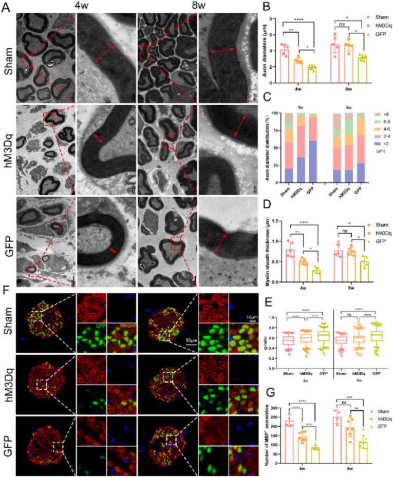
Results: We found that the AAV9-hM3Dq virus efficiently transduced motoneurons, and CNO administration robustly activated mature hM3Dq+ motoneurons in vivo. Chronic chemogenetic activation significantly enhanced the regeneration of spinal motoneurons injured by ventral root crush, accelerated axon extension, and improved axonal remyelination, resulting in increased axon size. This activation also facilitated the formation of new neuromuscular junctions (NMJs) in adult motoneurons and reduced muscle atrophy. Furthermore, it promoted electrophysiological recovery of the motor unit and improved overall motor function.

Conclusion: Chemogenetic activation of adult motoneurons can robustly enhances axon growth and mediate better behavioral recovery. These findings highlight the therapeutic potential of chemogenetic neuronal activation in promoting functional recovery following nerve injury.

The translational potential of this article: We have established a chronic chemogenetic method to activate hM3Dq+ motor neurons after brachial plexus injury, which accelerates axonal regeneration and enhances functional recovery. This strategy holds promise as a clinical therapeutic approach for treating nervous system injuries.

Keywords: Brachial plexus injury; Chemogenetic activation; Motor repair; Spinal motoneurons.


Conflict of interest statement

The authors declare that they have no known competing financial interests or personal relationships that could have appeared to influence the work reported in this paper. Lihua Zhou reports financial support was provided by Sun Yat-Sen University. Ke Zhong reports financial support was provided by Sun Yat-Sen Memorial Hospital. Reports a relationship with that includes:. Has patent pending to. If there are other authors, they declare that they have no known competing financial interests or personal relationships that could have appeared to influence the work reported in this paper.

Figures

Image 1
Graphical abstract
Fig. 1
Fig. 1
Intraspinal injection of AAV9- hM3Dq primarily transduces motoneurons and hM3Dq+ motoneurons were activated by CNO treatment. (A) AAV9-hM3Dq-GFP and AAV9-GFP was injected into the right C6 spinal ventral horn. One week later, C6 segment of spinal cord and its ventral root fibers were collected and immunostained for ChAT. Scale bar: 50 μm. (B) Quantification of percentages of GFP+ motor neurons, “0” indicates the injection point, while “-” indicates rostral side and “+” indicates caudal end. Data are expressed as mean ± SD (n = 3), analyzed by one-way ANOVA analysis of variance followed by Bonferroni's post hoc test. (C) Number of GFP+ ChAT+ merge ventral root fibers, data are expressed as mean ± SD (n = 3), analyzed by unpaired t test analysis with two-tailed P-value. (D) The data showed intraperitoneal CNO injection chemogenetically activated hM3Dq+ neurons in vivo. Experiment timeline is shown in the right panel. Very few neurons transduced with AAV-GFP expressed c-Fos while many hM3Dq+ neurons expressed c-Fos (showed as arrowheads). (E) Quantification of the percentage of GFP+ neurons that are also c-Fos+ after CNO administration. Data are expressed as mean ± SD, analyzed by unpaired t test analysis with two-tailed P-value (n = 6; ∗∗∗∗p < 0.0001, vs GFP+).
Fig. 2
Fig. 2
Chronic activation of adult hM3Dq+ motoneurons increases the number of FG retrograde labeled regenerative motoneurons in vivo (A, B) Representative images of FG-labeled ventral horn motoneurons at day 28 (A) and day 56 (B) post-injury. More FG-labeled motoneurons were observed in AAV9- hM3Dq transfected mice. Scale bar: 100 μm. (C) Quantitative analysis of FG-labeled motoneuron number in C6 spinal segment at day 28 and day 56 post-injury. Data are expressed as mean ± SD, analyzed by one-way ANOVA analysis of variance followed by the Tukey post hoc test. (n = 3; ∗∗∗p = 0.0003 vs. hM3Dq, ∗∗∗∗p < 0.0001 vs. GFP, ∗∗p = 0.0018 hM3Dq vs. GFP; ^ p = 0.0111 vs. hM3Dq, ^^ p = 0.0012 vs. GFP, nsp = 0.1092 hM3Dq vs. GFP).
Fig. 3
Fig. 3
Chronic chemogenetic activation of hM3Dq+ motoneurons in C6 spinal ventral horns promotes crush-injured axon regeneration (A) Representative images of GFP+- axons in horizontal sections of the 5 mm musculocutaneous nerve at day 28 and day 56 post-injury. Scale bars: 500 μm in upper row; 100 μm in lower row. (B) Representative images of GFP+ axons in cross sections of the injured distal musculocutaneous nerve at day 28 and day 56 post-injury. Scale bars: 100 μm. (C) Quantification of GFP+ axons at day 28 and day 56 post-injury. Data are expressed as mean ± SD, analyzed by one-way ANOVA analysis of variance followed by the Tukey post hoc test. (n = 8; ∗∗∗∗p < 0.0001 Sham vs. hM3Dq or GFP, ∗∗p = 0.0027 hM3Dq vs. GFP; ^^^^ p < 0.0001 Sham vs. hM3Dq or GFP, nsp = 0.2880 hM3Dq vs. GFP).
Fig. 4
Fig. 4
hM3Dq+ activation accelerates regenerating motor axons extending into the musculocutaneous nerve to innervate the biceps brachialis. (A, B) Representative images of GFP+ regenerating axons in the musculocutaneous nerve ending in the muscle fibers of the biceps brachialis and their enlarged image at day 28 (A) and day 56 (B) post-injury. Scale bars: 1 mm in the lower magnification on the left column; 200 μm in the higher magnification on the right column (M: muscle fibers of biceps; n: musculocutaneous nerve ending). (C) Quantification of numbers of GFP+ fibers. Data are expressed as mean ± SD (n = 5 in sham group at day 28, n = 8 in all other groups; ∗∗p = 0.0062 vs. hM3Dq, ∗∗∗∗p < 0.0001 vs. GFP, ∗p = 0.0406 hM3Dq vs. GFP), analyzed by one-way ANOVA analysis of variance followed by the Tukey post hoc test.
Fig. 5
Fig. 5
Chemogenetic activation of adult motoneurons improves axonal myelination and enhances cross section area of the regenerative axons after spinal ventral root injury (A) Electron micrographs of distal musculocutaneous nerve. Scale bars: 2 μm in the lower magnification on the left column; 200 nm in the higher magnification on the right column. (B) Quantification of axon diameter of distal musculocutaneous nerve. hM3Dq group show larger axon diameter than GFP (n = 5, ∗∗p = 0.0016 vs. hM3Dq, ∗∗∗∗p < 0.0001 vs. GFP, ∗P = 0.0353 hM3Dq vs. GFP; nsp = 0.9319 vs. hM3Dq, ^ p = 0.0127 vs. GFP, ^ p = 0.0242 hM3Dq vs. GFP). (C) Statistical analysis of the proportion of different axonal diameter in injured musculocutaneous nerve at day 28 and day 56 post-injury. (D) Quantification of myelin sheath thickness (n = 5, ∗∗p = 0.0057 vs. hM3Dq, ∗∗∗∗p < 0.0001 vs. GFP, ∗P = 0.0408 hM3Dq vs. GFP; nsp = 0.9606 vs. hM3Dq, ^ p = 0.0178 vs. GFP, ^ p = 0.0289 hM3Dq vs. GFP). (E) G-ratio analysis of distal musculocutaneous nerve. (More than 180 axons were measured in all group; ∗∗∗∗p < 0.0001; ^^^^ p < 0.0001, nsp = 0.0812 sham vs. hM3Dq). (F) Cross sections of distal musculocutaneous nerve (stained with MBP, red). Scale bars: 50 μm in the lower magnification on the left; 10 μm in the higher magnification on the right. (G) Number of MBP+ axons (n = 4 in sham groups, n = 7 in all other groups; ∗∗∗∗p < 0.0001 vs. hM3Dq, vs. GFP, ∗∗∗p = 0.0004 hM3Dq vs. GFP; nsp = 0.1135 vs. hM3Dq, ^^^ p = 0.0003 vs. GFP, ^^ p = 0.0075 hM3Dq vs. GFP). Data are expressed as mean ± SD, analyzed by one-way ANOVA analysis of variance followed by the Tukey post hoc test.
Fig. 6
Fig. 6
Chronic activation of hM3Dq+ motoneurons induced by CNO treatment facilitates reinnervation of NMJs at day 28 and day 56 post-injury (A, B) Representative images of NMJs labeled by α-bungarotoxin (red) and GFP (green) on the biceps brachialis at day 28 (A) and day 56 (B) post-injury. Scale bar: 100 μm. (C) Statistical analysis of the proportion of denervated, partially innervated, and fully innervated NMJs at day 28 and day 56 post-injury. (D) Total number of the NMJs on the biceps brachialis. Data are expressed as mean ± SD (n = 8, nsp = 0.1976 vs. hM3Dq ∗∗∗p = 0.0002 vs. GFP, ∗p = 0.0106 hM3Dq vs. GFP; nsp = 0.4545 vs. hM3Dq, ^^^ p = 0.0023 vs. GFP, ^ p = 0.0365 hM3Dq vs. GFP), analyzed by one-way ANOVA analysis of variance followed by the Tukey post hoc test.
Fig. 7
Fig. 7
Chronic chemogenetic neuronal activation reduces muscle atrophy and enhances EMG amplitude of re-innervated muscles (A) Representative images of bilateral biceps brachialis (the injured side (R) to the contralateral (L) side) and cross sections of the muscles stained with HE. Scale bars: 2 mm in left column; 100 μm in right column. (B) Representative images of EMG response and its individual action potentials. Scale bars: 5 mV in both sides, 500 ms and 2 ms in left and right side respectively. (C) Statistical analysis of the wet weight R/L ratios of biceps (n = 6; ∗∗∗p = 0.0004 vs. hM3Dq, ∗∗∗∗p < 0.0001 vs. GFP, ∗P = 0.0248 hM3Dq vs. GFP; nsp = 0.4839 vs. hM3Dq, ^^ p = 0.0039 vs. GFP, ^ p = 0.0405 hM3Dq vs. GFP). (D) Distribution of the cross-sectional areas of the muscle filaments in different groups. (E) Quantification of the cross-sectional areas of the muscle filaments (n = 6; ∗∗p = 0.0044 vs. hM3Dq, ∗∗∗∗p < 0.0001 vs. GFP, ∗P = 0.0345 hM3Dq vs. GFP; nsp = 0.8407 vs. hM3Dq, ^ p = 0.0245 vs. GFP, nsp = 0.0719 hM3Dq vs. GFP). (F) Averaged P–P value of the lesion side (n = 7 in sham groups, n = 8 in other groups; ∗∗∗p = 0.0001 vs. hM3Dq, ∗∗∗∗p < 0.0001 vs. GFP, ∗P = 0.0378 hM3Dq vs. GFP; ^^p = 0.0018 vs. hM3Dq, ^^^^ p < 0.0001 vs. GFP, ^ p = 0.0449 hM3Dq vs. GFP). Data are expressed as mean ± SD, analyzed by one-way ANOVA analysis of variance followed by the Tukey post hoc test.
Fig. 8
Fig. 8
Chronic activation of motoneurons enhances functional recovery of elbow flexion and walking ability post-injury (A) Positions of the forelimbs and corresponding scores (0–5) in the Terzis grooming test (TGT). (B) TGT scores pre- and post-injury (n = 12 in each group; ∗P < 0.05, ∗∗p < 0.01 hM3Dq vs. GFP). (C, D) Footprints of the upper limb at day 28 and day 56 post-surgery, RF: right front footprint, LF: left front footprint. (E–G) Percentage of R/L max contact area, R/L max intensity and R/L mean intensity from day 0 to day 56 post injury (n = 10 in each group; #P < 0.05 Sham vs. hM3Dq; ^P < 0.05 Sham vs. GFP; ∗P < 0.05 hM3Dq vs. GFP at the same time point). Data are expressed as the mean ± SD, analyzed by two-way ANOVA analysis of variance followed by the Tukey post hoc test.

Similar articles

Cited by

References

    1. Gutkowska O., Martynkiewicz J., Urban M., Gosk J. Brachial plexus injury after shoulder dislocation: a literature review. Neurosurg Rev. 2020;43(2):407–423. - PMC - PubMed
    1. Scheib J., Hoke A. Advances in peripheral nerve regeneration. Nat Rev Neurol. 2013;9(12):668–676. - PubMed
    1. Rasulić L., Savić A., Živković B., Vitošević F., Mićović M., Baščarević V., et al. Outcome after brachial plexus injury surgery and impact on quality of life. Acta Neurochir. 2017;159(7):1257–1264. - PubMed
    1. He Z., Jin Y. Intrinsic control of axon regeneration. Neuron. 2016;90(3):437–451. - PubMed
    1. Mahar M., Cavalli V. Intrinsic mechanisms of neuronal axon regeneration. Nat Rev Neurosci. 2018;19(6):323–337. - PMC - PubMed

LinkOut - more resources