. 2025 Apr 14;19(2):e70014.
doi: 10.1002/ccs3.70014. eCollection 2025 Jun.

Extracellular vesicle-LncRNA HOTAIR modulates esophageal cancer chemoresistance and immune microenvironment via miR-375/CDH2 pathway

Affiliations

Extracellular vesicle-LncRNA HOTAIR modulates esophageal cancer chemoresistance and immune microenvironment via miR-375/CDH2 pathway

Tayier Tuersong et al. J Cell Commun Signal. .

Abstract

Chemoresistance and immune evasion remain significant barriers to effective esophageal cancer (EC) treatment. This study explores the mechanistic role of extracellular vesicles (EVs) delivering LncRNA HOTAIR in modulating these processes. Using transcriptomic profiling, LncRNA HOTAIR was identified as a critical factor in EC progression. Its interaction with miR-375 was examined via luciferase reporter assays and RNA immunoprecipitation. Paclitaxel-resistant EC cells were treated with EVs containing HOTAIR, and the functional impact on proliferation, migration, invasion, and immune response was assessed through in vitro and in vivo models. LncRNA HOTAIR in EVs enhanced paclitaxel resistance by suppressing miR-375 and increasing CDH2 expression. Furthermore, HOTAIR promoted immune escape by upregulating PD-L1, impairing T-cell-mediated cytotoxicity. These changes were validated in patient-derived EC models. This study demonstrates that EV-LncRNA HOTAIR mediates chemoresistance and immune evasion in EC by targeting the miR-375/CDH2 axis. These findings provide a foundation for novel therapeutic interventions targeting EV-HOTAIR.

Keywords: CDH2; LncRNA HOTAIR; chemoresistance; esophageal cancer; extracellular vesicles; immune evasion; miR‐375.


Conflict of interest statement

The authors declare no conflicts of interest.

Figures
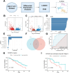
FIGURE 1
FIGURE 1
Screening of Key LncRNAs. (A) Flowchart for screening key LncRNAs; (B) volcano plot of differential expression in the GSE111011 dataset, showing significantly upregulated and downregulated genes (Normal: n = 7, EC: n = 7); (C) volcano plot of lncRNA expression in EC and normal tissues with blue and red indicating significantly downregulated and upregulated lncRNAs, respectively (Normal: n = 7, EC: n = 7); (D) SVM analysis showing the top 10 characteristic LncRNAs ranked by weight; (E) LASSO regression analysis showing selected LncRNAs and their corresponding regression coefficients; (F) Venn diagram of intersecting results from SVM and LASSO analyses; (G) ROC curves from TCGA database samples demonstrating the classification ability of intersecting LncRNAs (HOTAIR and TUSC7), with AUC values assessing model performance; (H) comparison of 3‐year truncated survival between high and low HOTAIR expression (high vs. low) in TCGA database samples; and (I) overall survival comparison between high and low HOTAIR expression (high vs. low) in Stage II samples from the TCGA database.
FIGURE 2
FIGURE 2
Regulatory Network of HOTAIR in EC and Expression Analysis of Its Target miRNA and mRNA. (A) Flowchart for screening key miRNAs and mRNAs; (B) miRNA‐target network of HOTAIR and its related miRNAs; (C) Venn diagram analysis of miRNAs identified from RNADisease database and selected miRNAs; (D) expression levels of hsa‐miR‐375 in EC and normal tissues from TCGA database (Normal: n = 13, EC: n = 184); (E) Venn diagram analysis of HOTAIR‐related mRNAs and hsa‐miR‐375 target mRNAs; (F) expression comparison of intersecting mRNAs between EC and normal tissues in TCGA database; (G) correlation analysis of hsa‐miR‐375 and CDH2 in TCGA database samples; (H) predicted binding sites of HOTAIR and CDH2 with miR‐375 using RNAhybrid tool; and (I) survival prediction capability of CDH2 expression for EC patients at different time points (1‐year, 3‐year, 5‐year) in TCGA database with blue, red, and green curves representing 1‐year, 3‐year, and 5‐year survival prediction curves, respectively.
FIGURE 3
FIGURE 3
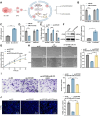
Validation and functional phenotyping of HOTAIR‐miR‐375/CDH2 axis interaction. (A) Schematic illustration of the effect of LncRNA HOTAIR on EC cell proliferation and invasion via miR‐375/CDH2 axis; (B) dual‐luciferase reporter assay showing the impact of HOTAIR overexpression on miR‐375 luciferase activity in KYSE30 cells; (C) RNA pull‐down assay evaluating HOTAIR‐mediated miR‐375 enrichment; (D) dual‐luciferase reporter assay detecting the effect of miR‐375 mimic transfection on CDH2 3′UTR luciferase activity; (E) RT‐qPCR detecting mRNA expression levels of HOTAIR, miR‐375, and CDH2; (F) WB detecting CDH2 protein expression; (G) CCK‐8 assay assessing cell proliferation; (H) wound healing assay evaluating cell migration (bar: 100 μm); (I) transwell invasion assay detecting cell invasion (bar: 50 μm); and (J) TUNEL staining detecting cell apoptosis rates (bar: 20 μm). Cell experiments were repeated three times. * indicates comparisons between groups, p < 0.05, **p < 0.01, ***p < 0.001, ****p < 0.001.
FIGURE 4
FIGURE 4
Characterization of EVs from KYSE30 and KYSE150 EC Cell lines and detection of HOTAIR. (A) Schematic diagram of EV isolation; (B) TEM observing the morphology of isolated EVs (bar: 200 nm); (C) NTA measuring the particle size distribution of isolated EVs; (D) WB detecting markers CD63, CD81, TSG101, and the negative marker Calnexin in isolated EVs; and (E) RT‐qPCR detecting HOTAIR expression levels in isolated EVs. Cell experiments were repeated three times. * indicates comparisons between groups, p < 0.05. CL: Cell lysate.
FIGURE 5
FIGURE 5
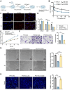
Effect of EVs‐mediated miR‐375/CDH2 axis on PTX resistance in KYSE30 cells. (A) Flowchart of constructing PTX‐resistant EC cell line KYSE30; (B) CCK‐8 assay evaluating the survival rate of resistant and sensitive cell lines after PTX treatment; (C) fluorescence microscopy showing the uptake efficiency of Dil‐labeled EVs by PTX‐resistant KYSE30 cells; (D) RT‐qPCR detecting mRNA expression levels of HOTAIR, miR‐375, and CDH2 in resistant cells; (E) CCK‐8 assay evaluating the survival rate of resistant cells in different treatment groups; (F) transwell migration assay assessing migration ability of resistant cells in different treatment groups (bar: 50 μm); (G) wound healing assay evaluating migration ability of resistant cells in different treatment groups (bar: 100 μm); and (H) TUNEL staining detecting apoptosis rates of resistant cells in different treatment groups. Cell experiments were repeated three times. * indicates comparisons between groups, p < 0.05, **p < 0.01, ***p < 0.001, ****p < 0.001.
FIGURE 6
FIGURE 6
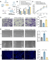
The effect of EV‐delivered LncRNA HOTAIR on PTX resistance in KYSE30 cells via the miR‐375/CDH2 axis. (A) Flowchart of EVs‐HOTAIR mediating the miR‐375/CDH2 axis and PTX resistance; (B) RT‐qPCR detecting expression levels of HOTAIR, CDH2, and miR‐375; (C) CCK‐8 assay evaluating the survival rate of KYSE30 cells in each group; (D) transwell migration assay assessing invasion ability of KYSE30 cells in each group; (E) wound healing assay evaluating migration ability of KYSE30 cells; and (F) TUNEL staining detecting apoptosis rates of KYSE30 cells in each group.
FIGURE 7
FIGURE 7
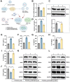
Study on the EVs‐mediated immune evasion mechanism. (A) Schematic diagram of the EVs‐mediated immune evasion mechanism in KYSE30; (B) RT‐qPCR detecting mRNA expression levels of PD‐L1 in different treatment groups of KYSE30 cells; (C) WB detecting protein expression levels of PD‐L1 in different treatment groups of KYSE30 cells; (D) CCK‐8 assay evaluating T cell‐mediated cytotoxicity against KYSE30 cells in different treatment groups; (E–J) ELISA detecting levels of TNF‐α, IFN‐γ, IL‐2, IL‐10, IL‐1β, and TGF‐β in co‐culture media of different treatment groups; and (K–L) WB analyzing activation levels of JAK/STAT and PI3K/AKT signaling pathways. Cell experiments were repeated three times. * indicates comparisons between groups, p < 0.05, **p < 0.01.
FIGURE 8
FIGURE 8
The effect of EV‐delivered LncRNA HOTAIR on immune evasion in KYSE30 cells via the miR‐375/CDH2 axis. (A) Experimental workflow of EVs‐LncRNA HOTAIR promoting immune evasion via the miR‐375/CDH2 axis; (B) RT‐qPCR detecting mRNA expression levels of PD‐L1 in different treatment groups of KYSE30 cells; (C) WB detecting protein expression levels of PD‐L1 in different treatment groups of KYSE30 cells; (D) CCK‐8 assay evaluating T cell‐mediated cytotoxicity against KYSE30 cells in different treatment groups; (E–J) ELISA detecting levels of TNF‐α, IFN‐γ, IL‐2, IL‐10, IL‐1β, and TGF‐β in co‐culture media of different treatment groups; and (K–L) WB analyzing activation levels of JAK/STAT and PI3K/AKT signaling pathways. Cell experiments were repeated three times. * indicates comparisons between groups, p < 0.05, **p < 0.01. Group 1 represents PTX + EVs‐si‐NC, Group 2 represents PTX + EVs‐si‐HOTAIR + oe‐NC, and Group 3 represents PTX + EVs‐si‐HOTAIR + oe‐CDH2.

Similar articles

Cited by

References

    1. Wang, Y. , Yang W., Wang Q., and Zhou Y.. 2023. “Mechanisms of Esophageal Cancer Metastasis and Treatment Progress.” Frontiers in Immunology 14. 10.3389/fimmu.2023.1206504. - DOI - PMC - PubMed
    1. Li, R. , Huang B., Tian H., and Sun Z.. 2023. “Immune Evasion in Esophageal Squamous Cell Cancer: From the Perspective of Tumor Microenvironment.” Frontiers in Oncology 12. 10.3389/fonc.2022.1096717. - DOI - PMC - PubMed
    1. Zhou, C. , Fan N., Liu F., Fang N., Plum P. S., Thieme R., Gockel I., et al. 2020. “Linking Cancer Stem Cell Plasticity to Therapeutic Resistance‐Mechanism and Novel Therapeutic Strategies in Esophageal Cancer.” Cells 9(6): 1481. 10.3390/cells9061481. - DOI - PMC - PubMed
    1. Ning, X.‐Y. , Ma J.‐H., He W., and Ma J.‐T.. 2023. “Role of Exosomes in Metastasis and Therapeutic Resistance in Esophageal Cancer.” World Journal of Gastroenterology 29(42): 5699–5715. 10.3748/wjg.v29.i42.5699. - DOI - PMC - PubMed
    1. Mao, C. , Zeng X., Zhang C., Yang Y., Xiao X., Luan S., Zhang Y., and Yuan Y.. 2021. “Mechanisms of Pharmaceutical Therapy and Drug Resistance in Esophageal Cancer.” Frontiers in Cell and Developmental Biology 9. 10.3389/fcell.2021.612451. - DOI - PMC - PubMed