. 2025 Apr 1:13:1565262.
doi: 10.3389/fcell.2025.1565262. eCollection 2025.

Chloride intracellular channel (CLIC) protein function in S1P-induced Rac1 activation requires membrane localization of the C-terminus, but not thiol-transferase nor ion channel activities

Affiliations

Chloride intracellular channel (CLIC) protein function in S1P-induced Rac1 activation requires membrane localization of the C-terminus, but not thiol-transferase nor ion channel activities

De Yu Mao et al. Front Cell Dev Biol. .

Abstract

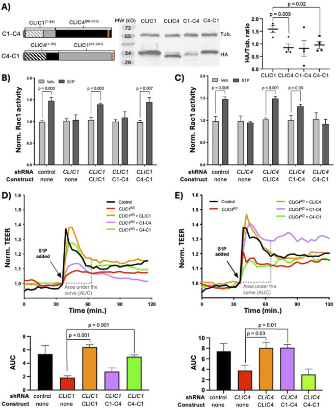
We have established a novel and evolutionarily-conserved function for chloride intracellular channel proteins (CLICs) in regulating Rho/Rac GTPases downstream of G protein-coupled receptors (GPCRs). Endothelial CLIC1 and CLIC4 are rapidly and transiently re-localized from the cytoplasm to the plasma membrane in response to the GPCR ligand sphingosine-1-phosphate (S1P), and both CLICs are required to activate Rac1 in response to S1P, but how they perform this function remains unknown. Biochemical studies suggest that CLICs act as non-specific ion channels and/or as glutathione-S-transferases, dependent on N-terminal features, in vitro. Here we investigate CLIC functional domains and membrane localization requirements for their function in S1P-mediated Rac1 signaling. Structure-function analyses of CLIC function in endothelial cells demonstrate that CLIC1 and CLIC4-specific functions reside at their C-termini, and that the CLIC4 N-terminus encodes determinants required for S1P-induced re-localization to the plasma membrane but is dispensable for S1P-induced Rac1 activation when the C-terminus is localized to the plasma membrane via a heterologous signal. Our results demonstrate that the postulated ion channel and thiol-transferase activities of CLICs are not required for Rac1 activation and suggests that sequences in the CLIC C-termini are critical for this function. Given the importance of S1P signaling in vascular biology and disease, our work establishes a platform to further our understanding of the membrane-localized proteins required to link GPCR activity to Rho/Rac regulation.

Keywords: CLIC1; CLIC4; Rac1; RhoA; S1P receptor; endothelium; sphingosine-1-phosphate.


Conflict of interest statement

The authors declare that the research was conducted in the absence of any commercial or financial relationships that could be construed as a potential conflict of interest.

Figures

FIGURE 1
FIGURE 1
CLIC1 and CLIC4 in endothelial S1P signaling, and CLIC sequence and structural features relevant to this study. (A) description of shared and unique functions for CLIC1 and CLIC4 in endothelial S1P signaling, as defined in Mao et al. (2021). (B) Protein sequence alignment of C. elegans EXC-4, and human CLIC1 and CLIC4 indicating regions of amino-acid identity (dark grey shading) and similarity (light grey shading). Conserved beta sheets (yellow arrows) and alpha helices (pink rounded boxes). The N-terminal putative transmembrane domain (PTMD) and conserved thioredoxin fold are highlighted. Note that the N-terminal cysteine required for thiol-reductases activity (red), the cholesterol-binding GxxxG motif (brown), and C-terminal nuclear localization signal (orange) found in human CLIC1 and four are not present in C. elegans EXC-4, while the C. elegans protein has a C-terminal extension with additional alpha helices (α10, α11). (C) cartoon representation of HA-tagged full-length CLIC1 and CLIC4 constructs. The C-terminal fragments swapped in chimeric constructs (Figure 2) are denoted in blue, and the truncated (∆PTMD)CLIC4 construct (Figures 3, 4) is denoted in purple [the starting protein sequence for chimeric and truncated constructs are shown as blue and purple arrows, respectively, in panel (B)].
FIGURE 2
FIGURE 2
CLIC1 and CLIC4-specific functions in S1P signaling are encoded by their C-termini. (A) cartoon representation of the C1-C4 and C4-C1 chimeras, and western blot analysis and quantification (n = 4) of their expression in HUVEC (see Methods). In panels (B) and (C) active Rac1 measured by G-LISA (see Methods). Here, and in subsequent figures showing G-LISA results, each bar represents the mean of n ≥ 3 replicates, and error bars represent standard deviation (SD). Significance between vehicle control (Veh.) and S1P-treated cells was calculated via two-tailed unpaired t-tests. (B) full-length CLIC1 and the C4-C1 chimera rescue the Rac1 activation defect caused by CLIC1 KD , while (C) full-length CLIC4 and the C1-C4 chimera rescue the Rac1 activation defect caused by CLIC4 KD . In panels (D, E), and in subsequent figures showing TEER results, each trace represents the mean trans-endothelial electrical resistance (TEER) measurement of n ≥ 3 samples, normalized to the TEER value at time of S1P addition (see Methods). To quantify TEER results, the area under the curve (AUC) ± standard error of the mean (SEM) from the time of S1P addition (∼30min after beginning of resistance measurements) until time of maximal recovery to baseline (∼30 min after S1P addition in controls) was calculated for each condition (see Methods), and results are shown in the accompanying bar graphs. Significance was calculated using unpaired two-tailed t tests. (D) full-length CLIC1 and the C4-C1 chimera rescue the barrier defect caused by CLIC1 KD , while (E) full-length CLIC4 and the C1-C4 chimera rescue the TEER defect caused by CLIC4 KD .
FIGURE 3
FIGURE 3
The N-terminal putative transmembrane domain (PTMD) is required for CLIC4 re-localization and Rac1 activation in response to S1P. (A) cartoon representation of the HA-tagged (∆PTMD)CLIC4 construct, and western blot analysis and quantification of its expression in HUVEC (see Methods), as compared to full-length HA-CLIC4. (B) Immunofluorescence of full-length HA-tagged CLIC4 (top) and (∆PTMD)CLIC4, and their localization after treatment with S1P. Yellow arrows denote PM localization. Scale bar represents 50 µm in all panels. (C) the (∆PTMD)CLIC4 construct does not rescues the Rac1 activation defect caused by CLIC4 KD , as assessed by G-LISA. (D) the (∆PTMD)CLIC4 construct does not rescue the TEER defect caused by CLIC4 KD .
FIGURE 4
FIGURE 4
Membrane localized CLIC4 C-terminus is sufficient to promote Rac1 activation in response to S1P. (A) cartoon representation of HA-tagged full-length myristylated (myr) CLIC4 and myr-(∆PTMD)CLIC4, lacking the N-terminal putative transmembrane domain. Western blot analysis and quantification of expression in HUVEC (see Methods), shows lower expression of myr-(∆PTMD)CLIC4 as compared to full-length myr-CLIC4. (B) Immunofluorescence showing accumulation of HA-tagged full-length CLIC4 (first column), myr-CLIC4 (second column) and myr-(∆PTMD)CLIC4 (third column) at steady state (no S1P). Note that both myristylated constructs display accumulation at the membrane (yellow arrows), and decreased nuclear accumulation (cyan asterisks) when compared to wildtype full-length CLIC4. Scale bar represents 50 µm in all panels. (C) both the full-length myr-CLIC4 and the myr-(∆PTMD)CLIC4 constructs rescue the Rac1 activation defect caused by CLIC4 KD , as assessed by G-LISA. (D) both the full-length myr-CLIC4 and the myr-(∆PTMD)CLIC4 constructs rescue the TEER defect caused by CLIC4 KD . Dashed lines and bars (Control, CLIC4 KD , and CLIC4 KD + CLIC4) are the same data shown in Figure 3D.

Update of

References

    1. Alghalayini A., Hossain K. R., Moghaddasi S., Turkewitz D. R., D'Amario C., Wallach M., et al. (2023). In Vitro enzymatic studies reveal pH and temperature sensitive properties of the CLIC proteins. Biomolecules 13 (9), 1394. 10.3390/biom13091394 - DOI - PMC - PubMed
    1. Al Khamici H., Brown L. J., Hossain K. R., Hudson A. L., Sinclair-Burton A. A., Ng J. P., et al. (2015). Members of the chloride intracellular ion channel protein family demonstrate glutaredoxin-like enzymatic activity. PLoS One 10 (1), e115699. 10.1371/journal.pone.0115699 - DOI - PMC - PubMed
    1. Al Khamici H., Hossain K. R., Cornell B. A., Valenzuela S. M. (2016). Investigating sterol and redox regulation of the ion channel activity of CLIC1 using tethered bilayer membranes. Membr. (Basel) 6 (4), 51. 10.3390/membranes6040051 - DOI - PMC - PubMed
    1. Arena A. F., Escudero J., Shaye D. D. (2022). A metazoan-specific C-terminal motif in EXC-4 and Gα-Rho/Rac signaling regulate cell outgrowth during tubulogenesis in C. elegans . Development 149 (24), dev200748. 10.1242/dev.200748 - DOI - PMC - PubMed
    1. Argenzio E., Moolenaar W. H. (2016). Emerging biological roles of Cl-intracellular channel proteins. J. Cell Sci. 129 (22), 4165–4174. 10.1242/jcs.189795 - DOI - PubMed

LinkOut - more resources