. 2025 Apr 1;15(4):3285-3297.
doi: 10.21037/qims-24-2077. Epub 2025 Mar 28.

Spectral computed tomography parameters for predicting vessels encapsulating tumor clusters (VETC) pattern in hepatocellular carcinoma: a pilot study

Affiliations

Spectral computed tomography parameters for predicting vessels encapsulating tumor clusters (VETC) pattern in hepatocellular carcinoma: a pilot study

Yongquan Yu et al. Quant Imaging Med Surg. .

Abstract

Background: Vessels encapsulating tumor clusters (VETC) is a novel microvascular pattern associated with poor prognosis in patients with hepatocellular carcinoma (HCC). Reliable preoperative predictors of VETC may substantially improve prognostic outcomes. This study aimed to evaluate the predictive value of multiparameter spectral computed tomography (CT) in identifying VETC pattern in HCC.

Methods: This retrospective analysis included 50 patients with histopathologically confirmed HCC who underwent preoperative abdominal spectral CT and CD34 immunohistochemical staining. VETC(+) was defined as a visible vessel-encapsulating tumor cluster occupying ≥5% of the tumor area. Patients were divided into VETC(+) (n=21) and VETC(-) (n=29) groups. Eight qualitative imaging features, including tumor size, intratumor vascularity, nonrim arterial phase (AP) hyperenhancement, nonperipheral portal phase (PP) washout, well-defined capsule, nonsmooth tumor margin, intratumoral necrosis, and AP hypovascular component, were assessed via 40-keV virtual monoenergetic images (VMIs). Quantitative spectral parameters, including iodine concentration (IC), normalized IC (NIC), effective atomic number (Zeff), and energy spectrum curve slope (λ), were measured in both the PP and equilibrium phase (EP). Multivariate logistic regression analysis was performed to identify independent risk factors for VETC pattern, receiver operating characteristic (ROC) analysis was used to evaluate the diagnostic performance, and the Kaplan-Meier method was used to assess recurrence-free survival (RFS).

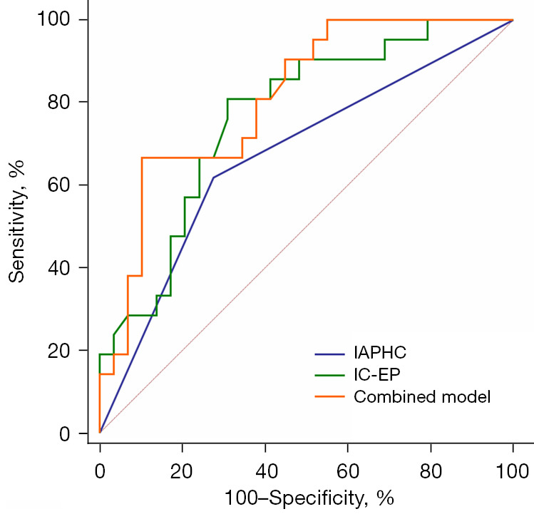
Results: Significant differences were found between the VETC(+) and VETC(-) groups in alpha-fetoprotein level, intratumor AP hypovascular component (IAPHC), and portal- and equilibrium-phase spectral parameters (IC, Zeff, and λ; all P values <0.05). Multivariate logistic regression identified the IAPHC and EP IC as independent VETC predictors of VETC patter [odds ratio (OR) =4.149 and OR =12.724, respectively]. The combined model demonstrated superior diagnostic performance (AUC =0.810) compared to the individual parameters (IAPHC: AUC =0.672; EP IC: AUC =0.766), achieving a 66.67% sensitivity and an 89.66% specificity. Kaplan-Meier survival analysis indicated a shorter RFS in the VETC(+) group than in the VETC(-) group (P=0.02).

Conclusions: IAPHC and EP IC derived from spectral CT hold significant potential for VETC prediction in HCC. The application of a combined model enhances diagnostic efficiency.

Keywords: Hepatocellular carcinoma (HCC); differential diagnosis; spectral computed tomography (spectral CT); vessels encapsulating tumor clusters (VETC).


Conflict of interest statement

Conflicts of Interest: All authors have completed the ICMJE uniform disclosure form (available at https://qims.amegroups.com/article/view/10.21037/qims-24-2077/coif). X.C. is an employee of Philips Healthcare, the manufacturer of the CT system used in this study. The other authors have no conflicts of interest to declare.

Figures

Figure 1
Figure 1
Flowchart of participant enrollment. AFP, alpha-fetoprotein; CEA, carcinoembryonic antigen; CT, computed tomography; HCC, hepatocellular carcinoma; PIVKA-II, protein-induced by vitamin K absence or antagonist-II; VETC, vessels encapsulating tumor clusters.
Figure 2
Figure 2
Comparison of AP CTConvention and CT40keV-VMI images in VETC-positive/negative HCCs (red boxes indicate lesions). (A,B) In VETC-positive HCC, the tumor exhibited uneven and diffuse enhancement in the AP. The CT40keV-VMI image revealed an IAPHC and necrotic regions within the tumor, which were more prominent as compared to the CTConvention image. (C,D) In VETC-negative HCC, the tumor showed mild and homogeneous enhancement in the AP. The lesion appeared less distinct on the CTConvention image, whereas it was clearly depicted on the CT40keV-VMI image. AP, arterial phase; CT, computed tomography; CT40keV-VMI, 40-keV virtual monoenergetic images CT; CTConvention, conventional CT; HCC, hepatocellular carcinoma; IAPHC, intratumor arterial phase hypovascular component; VETC, vessels encapsulating tumor clusters.
Figure 3
Figure 3
Column charts showing the IC-PP (A), NIC-PP (B), Zeff-PP (C), λ-PP (D), IC-EP (E), NIC-EP (F), Zeff-EP (G), and λ-EP (H) of VETC(+) and VETC(−) HCCs. *, P<0.05; ns, not significant (P>0.05). HCC, hepatocellular carcinoma; IC-EP, iodine concentration in the equilibrium phase; IC-PP, iodine concentration in the portal phase; λ-EP, energy spectrum curve slope of the equilibrium phase; λ-PP, energy spectrum curve slope of the portal phase; NIC-EP, normalized iodine concentration in the equilibrium phase; NIC-PP, normalized iodine concentration in the portal phase; VETC, vessels encapsulating tumor clusters; Zeff-EP, effective atomic number in the equilibrium phase; Zeff-PP, effective atomic number in the portal phase.
Figure 4
Figure 4
Representative histopathological and radiological images of an HCC case with VETC (59-year-old female). (A-D) Conventional image, IC map, Zeff map, and spectral curve of the PP (red arrows indicate the lesions), respectively. (E-H) Conventional image, IC map, Zeff map, and spectral curve of the EP (red arrows indicate the lesions), respectively. (I,J) Typical VETC pattern tumor cluster captured by a web-like vascular network [(I) hematoxylin and eosin staining, magnification 100×; (J) CD34 immunohistochemical staining, magnification 100×]. AP, arterial phase; EP, equilibrium phase; HCC, hepatocellular carcinoma; HU, Hounsfield units; IC, iodine concentration; ROI, region of interest; VETC, vessels encapsulating tumor clusters; Zeff, effective atomic number.
Figure 5
Figure 5
Representative histopathological and radiological images of an HCC case without VETC (67-year-old male). (A-D) Conventional image, IC map, Zeff map, and spectral curve of the PP (red arrows indicate the lesions), respectively. (E-H) Conventional image, IC map, Zeff map, and spectral curve of the EP (red arrows indicate the lesions), respectively. (I,J) A classical capillary vascular pattern [(I) hematoxylin and eosin staining, magnification 100×; (J) CD34 immunohistochemical staining, magnification 100×]. AP, arterial phase; EP, equilibrium phase; HCC, hepatocellular carcinoma; HU, Hounsfield units; IC, iodine concentration; ROI, region of interest; VETC, vessels encapsulating tumor clusters; Zeff, effective atomic number.
Figure 6
Figure 6
ROC curves of the IAPHC, IC-EP, and the combined model for predicting the VETC pattern. IC-EP, iodine concentration in the equilibrium phase; IAPHC, intratumor arterial phase hypovascular component; ROC, receiver operating characteristic; VETC, vessels encapsulating tumor clusters.
Figure 7
Figure 7
Kaplan-Meier curve for postoperative RFS according to the histological VETC pattern. Patients in the VETC(+) group exhibited poorer RFS than did those in the VETC(−) group (P=0.02). RFS, recurrence-free survival; VETC, vessels encapsulating tumor clusters.

Similar articles

References

    1. Sung H, Ferlay J, Siegel RL, Laversanne M, Soerjomataram I, Jemal A, Bray F. Global Cancer Statistics 2020: GLOBOCAN Estimates of Incidence and Mortality Worldwide for 36 Cancers in 185 Countries. CA Cancer J Clin 2021;71:209-49. 10.3322/caac.21660 - DOI - PubMed
    1. Sun Y, Wu L, Zhong Y, Zhou K, Hou Y, Wang Z, et al. Single-cell landscape of the ecosystem in early-relapse hepatocellular carcinoma. Cell 2021;184:404-421.e16. 10.1016/j.cell.2020.11.041 - DOI - PubMed
    1. Hepatocellular carcinoma. Nat Rev Dis Primers 2021;7:7. 10.1038/s41572-021-00245-6 - DOI - PubMed
    1. Fang JH, Zhou HC, Zhang C, Shang LR, Zhang L, Xu J, Zheng L, Yuan Y, Guo RP, Jia WH, Yun JP, Chen MS, Zhang Y, Zhuang SM. A novel vascular pattern promotes metastasis of hepatocellular carcinoma in an epithelial-mesenchymal transition-independent manner. Hepatology 2015;62:452-65. 10.1002/hep.27760 - DOI - PubMed
    1. Li Z, Song W, Zhang J, Li Q, Song Z, Ren X, Wen Y, Li X, Yao H, Gao Y, Tang Z. Identification of vessels encapsulating tumor clusters in solitary hepatocellular carcinoma via imaging biomarkers in preoperative contrast-enhanced magnetic resonance imaging. Quant Imaging Med Surg 2024;14:8586-600. 10.21037/qims-24-315 - DOI - PMC - PubMed

LinkOut - more resources