

doi: 10.21037/qims-24-2420.
Epub 2025 Mar 28.
Prenatal diagnosis of coronary artery to right ventricle fistula: a case description
Affiliations
- PMID: 40235775
- PMCID: PMC11994515
- DOI: 10.21037/qims-24-2420
Item in Clipboard
Prenatal diagnosis of coronary artery to right ventricle fistula: a case description
Quant Imaging Med Surg.
.
No abstract available
Conflict of interest statement
Conflicts of Interest: All authors have completed the ICMJE uniform disclosure form (available at https://qims.amegroups.com/article/view/10.21037/qims-24-2420/coif). L.Y. reports receiving funding from the Lanzhou Talent Innovation and Entrepreneurship Project (No. 2023-RC-23). The other authors have no conflicts of interest to declare.
Figures
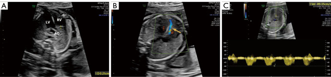
Two-dimensional and Doppler ultrasound imaging was conducted for critical assessment of the fetal coronary artery and abnormal shunt hemodynamics in the fetal heart. (A) Fetal heart two-dimensional ultrasound was used to measure the diameter of the coronary artery. (B) Color Doppler indicated a continuous blood flow signal from the apex toward the tricuspid valve (arrow). (C) Measurement of blood flow velocity. AO, aorta; LV, left ventricle; RV, right ventricle.
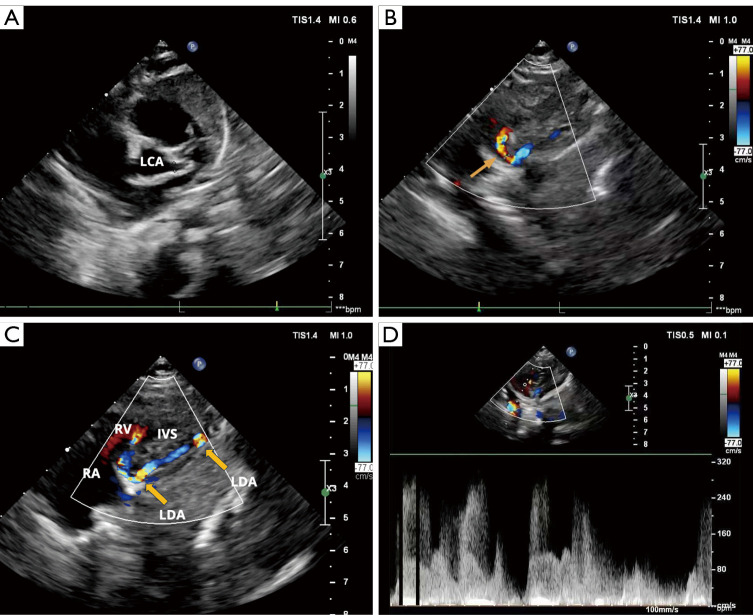
Postnatal two-dimensional and color Doppler echocardiographic images of the infant provided key measurements of the coronary arteries and demonstrated abnormal shunt flow. (A) Inner diameter of the left coronary artery. (B) Inner diameter of the left coronary artery fistula into the right ventricular fistula opening (arrow). (C) Color Doppler indicated that the left anterior descending artery reached the apex and then looped along the posterior interventricular septum to the posterior atrioventricular groove, showing bright turbulent flow at the connection sites with the right ventricle at the apical and atrioventricular groove regions. (D) The continuous shunt flow velocity was approximately 300 cm/s. IVS, interventricular septum; LCA, left coronary artery; LDA, left descending artery; RA, right atrium; RV, right ventricle.
Similar articles
-
An unusual case of prenatal diagnosis of right coronary artery to right ventricle fistula with HD-flow render mode and spatiotemporal image correlation (STIC).Echocardiography. 2020 Jul;37(7):1105-1108. doi: 10.1111/echo.14693. Epub 2020 Jun 28. Echocardiography. 2020. PMID: 32594574
-
Prenatal diagnosis of fetal isolated right coronary artery to left ventricle fistula.Echocardiography. 2019 May;36(5):1009-1013. doi: 10.1111/echo.14340. Epub 2019 Apr 19. Echocardiography. 2019. PMID: 31002180
-
A large hemodynamically significant right coronary artery fistula to right ventricle: prenatal detection and progression.Echocardiography. 2012 Aug;29(7):E173-5. doi: 10.1111/j.1540-8175.2012.01682.x. Epub 2012 Apr 4. Echocardiography. 2012. PMID: 22486425
-
[Single left coronary artery with a fistula to the right ventricle: report of a case with successful closure].Kyobu Geka. 1989 Apr;42(4):325-9. Kyobu Geka. 1989. PMID: 2671456 Review. Japanese.
-
Prenatal diagnosis of isolated coronary artery fistula: systematic review, analysis of perinatal prognostic factors and case report.J Matern Fetal Neonatal Med. 2023 Dec;36(1):2206938. doi: 10.1080/14767058.2023.2206938. J Matern Fetal Neonatal Med. 2023. PMID: 37121905
References
-
- Gómez-Arriaga PI, Escribano D, Gómez-Montes E, Villalaín C, Mendoza A, Galindo A. Prenatal diagnosis of isolated coronary artery fistula: systematic review, analysis of perinatal prognostic factors and case report. J Matern Fetal Neonatal Med 2023;36:2206938. 10.1080/14767058.2023.2206938 - DOI - PubMed
LinkOut - more resources
Full Text Sources