

Diagnostic accuracy of deep learning for the invasiveness assessment of ground-glass nodules with fine segmentation: a systematic review and meta-analysis
- PMID: 40235789
- PMCID: PMC11994546
- DOI: 10.21037/qims-24-1839
Diagnostic accuracy of deep learning for the invasiveness assessment of ground-glass nodules with fine segmentation: a systematic review and meta-analysis
Abstract
Background: Accurate recognition of invasive lung adenocarcinoma (IAC) presenting as ground-glass nodules (GGNs) is crucial for guiding clinical decision-making and timely surgical intervention. This study aimed to systematically evaluate the diagnostic accuracy of deep learning (DL) models via fine nodule segmentation in assessing the invasiveness of lung adenocarcinoma.
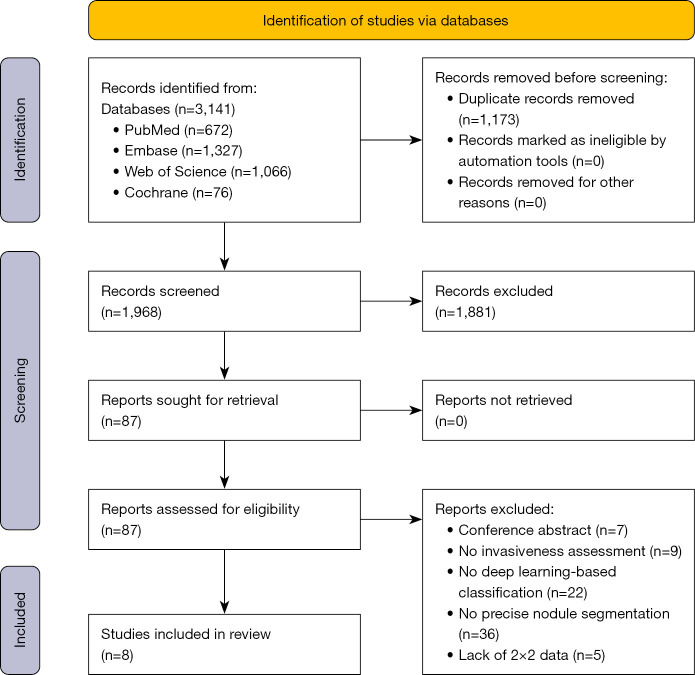
Methods: Literature from the inception of the PubMed, Embase, Cochrane Library, and Web of Science databases was searched. Studies related to DL and nodule segmentation in diagnosing IAC were evaluated and included. Titles and abstracts were screened, and the Quality Assessment of Diagnostic Accuracy Studies 2 was used to assess the quality of the selected studies. The Grading of Recommendations, Assessment, Development, and Evaluation (GRADE) criteria of diagnostic tests were used to assess the certainty of evidence.
Results: Eight studies involving 5,281 nodules and 4,676 patients were included and analyzed. Meta-analysis showed that the combined sensitivity of DL for the diagnosis of IAC was 0.81 [95% confidence interval (CI): 0.73-0.87], while the specificity was 0.86 (95% CI: 0.80-0.90). The area under the summary receiver operating characteristic (SROC) curve was 0.90 (95% CI: 0.88-0.93), but the overall quality of the evidence was suboptimal.
Conclusions: DL and nodule segmentation demonstrated high accuracy in assessing lung adenocarcinoma invasiveness, but the certainty of the associated evidence was low. More large-scale, multicenter, high-quality diagnostic accuracy studies are needed to validate the performance and usefulness of DL in the assessment of lung adenocarcinoma invasiveness.
Keywords: Artificial intelligence (AI); computed tomography (CT); deep learning (DL); invasive lung adenocarcinoma (IAC); pulmonary nodules.
Copyright © 2025 AME Publishing Company. All rights reserved.
Conflict of interest statement
Conflicts of Interest: All authors have completed the ICMJE uniform disclosure form (available at https://qims.amegroups.com/article/view/10.21037/qims-24-1839/coif). The authors have no conflicts of interest to declare.
Figures


Similar articles
-
The use of the mean computed-tomography value to predict the invasiveness of ground-glass nodules: A meta-analysis.Asian J Surg. 2023 Feb;46(2):677-682. doi: 10.1016/j.asjsur.2022.07.031. Epub 2022 Jul 19. Asian J Surg. 2023. PMID: 35864044 Review.
-
Lung-PNet: An Automated Deep Learning Model for the Diagnosis of Invasive Adenocarcinoma in Pure Ground-Glass Nodules on Chest CT.AJR Am J Roentgenol. 2024 Jan;222(1):e2329674. doi: 10.2214/AJR.23.29674. Epub 2023 Jul 26. AJR Am J Roentgenol. 2024. PMID: 37493322
-
Development and validation of a risk prediction model for invasiveness of pure ground-glass nodules based on a systematic review and meta-analysis.BMC Med Imaging. 2024 Jun 17;24(1):149. doi: 10.1186/s12880-024-01313-5. BMC Med Imaging. 2024. PMID: 38886695 Free PMC article.
-
Lung Adenocarcinoma Manifesting as Ground-Glass Opacity Nodules 3 cm or Smaller: Evaluation With Combined High-Resolution CT and PET/CT Modality.AJR Am J Roentgenol. 2019 Nov;213(5):W236-W245. doi: 10.2214/AJR.19.21382. Epub 2019 Jul 30. AJR Am J Roentgenol. 2019. PMID: 31361533
-
Exploring the optimal threshold of 3D consolidation tumor ratio value segmentation based on artificial intelligence for predicting the invasive degree of T1 lung adenocarcinoma.Quant Imaging Med Surg. 2024 Dec 5;14(12):8988-8998. doi: 10.21037/qims-24-1328. Epub 2024 Nov 29. Quant Imaging Med Surg. 2024. PMID: 39698672 Free PMC article.
References
-
- Church TR, Black WC, Aberle DR, Berg CD, Clingan KL, Duan F, Fagerstrom RM, Gareen IF, Gierada DS, Jones GC, Mahon I, Marcus PM, Sicks JD, Jain A, Baum S. Results of initial low-dose computed tomographic screening for lung cancer. N Engl J Med 2013;368:1980-91. 10.1056/NEJMoa1209120 - DOI - PMC - PubMed
LinkOut - more resources
Full Text Sources