

Primary hydatid of posterior thigh and popliteal fossa: A rare case report
- PMID: 40235839
- PMCID: PMC11999065
- DOI: 10.1016/j.jpra.2024.11.008
Primary hydatid of posterior thigh and popliteal fossa: A rare case report
Abstract
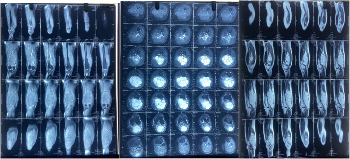
Hydatid disease of skeletal muscle is a rare entity. Liver followed by lung are the most common sites of hydatid cyst. Incidence of unusual sites which include spleen, kidney, mesentry, ovary, brain, skeletal system is 7,8 %. Skeletal muscle has the lowest incidence of primary hydatid among the unusual sites. Here we report a young female with rare primary hydatid disease of left posterior thigh extending into popliteal fossa.
Keywords: Cyst; Echinococus granulosus; Hydatid and rim sign; Popliteal; Skeletal muscle.
© 2025 The Authors. Published by Elsevier Ltd on behalf of British Association of Plastic, Reconstructive and Aesthetic Surgeons.
Conflict of interest statement
Authors declare that they have no conflict of interest. Proper consent for the photographs has been taken from the patient.
Figures


References
-
- White C., Jr, Weller P.F. In: Harrison's Principles of Internal Medicine. 15th edition. Braunwald E, Fauci AS, Kasper DL, Longo DL, Jameson JL, editors. McGraw Hill; 2001. Echinococcosis; p. 1250.
-
- Arambulo P., 3rd Public health importance of cystic echinococcosis in Latin America. Acta Trop. 1997;67:113–124. - PubMed
-
- Alintas N. Cystic and alveolar echinococcosis in Turkey. Ann Trop Med Parasitol. 1998;92:637–642. - PubMed
-
- Eckert J., Thompson R.C. Echinococcus strains in Europe: a review. Trop Med Parasitol. 1988;39:1–8. - PubMed
-
- Tatari H., Baran O., Anlidag T., et al. Primary intramuscular hydatidosis of supraspinatus muscle. Arch Orthop Trauma Surg. 2001;121:93–99. - PubMed
Publication types
LinkOut - more resources
Full Text Sources
