

Pancreatic neuroendocrine neoplasms coexisting with biliary intraductal papillary mucinous neoplasm: A case report and review of literature
- PMID: 40235901
- PMCID: PMC11995319
- DOI: 10.4251/wjgo.v17.i4.100497
Pancreatic neuroendocrine neoplasms coexisting with biliary intraductal papillary mucinous neoplasm: A case report and review of literature
Abstract
Background: Pancreatic neuroendocrine neoplasms (pNENs) are rare, heterogeneous tumors accounting for 1%-2% of pancreatic tumors, with significant malignant potential. Intraductal papillary mucinous neoplasm of the bile duct (IPMN-B) is a rare precancerous lesion in the bile duct system, with potential for malignancy. The combination of pNENs and IPMN-B is exceptionally rare and often leads to misdiagnosis. This study aims to report a rare case of pNENs combined with IPMN-B treated at Yanbian University Hospital to improve understanding and management of this unusual tumor combination.
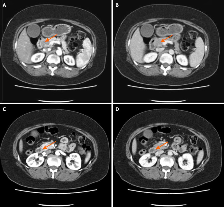
Case summary: We retrospectively analyzed a case from Yanbian University Hospital. We reviewed clinical records, imaging findings, endoscopic retrograde cholangiopancreatography, surgical exploration, and histopathological examination. The patient was diagnosed with pNENs and IPMN-B. Surgical treatment was performed, with follow-up showing effective management and no significant recurrence.
Conclusion: This case represents the first report of pNENs combined with IPMN-B. It highlights the need for thorough diagnostic evaluation to prevent misdiagnosis and improve treatment strategies.
Keywords: Case report; Endoscopic retrograde cholangiopancreatography; Histopathology; Intraductal papillary mucinous neoplasm of the bile duct; Malignant potential; Pancreatic neuroendocrine neoplasms.
©The Author(s) 2025. Published by Baishideng Publishing Group Inc. All rights reserved.
Conflict of interest statement
Conflict-of-interest statement: All the authors report no relevant conflicts of interest for this article.
Figures


References
-
- Pitman MB, Centeno BA, Reid MD, Siddiqui MT, Layfield LJ, Perez-Machado M, Weynand B, Stelow EB, Lozano MD, Fukushima N, Cree IA, Mehrotra R, Schmitt FC, Field AS. The World Health Organization Reporting System for Pancreaticobiliary Cytopathology. Acta Cytol. 2023;67:304–320. - PubMed
-
- Pitman MB, Centeno BA, Reid MD, Saeig M, Siddiqui MT, Layfield LJ, Perez-Machado M, Weynand B, Stelow EB, Lozano MD, Fukushima N, Cree IA, Mehrotra R, Schmitt FC, Field AS. A brief review of the WHO reporting system for pancreaticobiliary cytopathology. J Am Soc Cytopathol. 2023;12:243–250. - PubMed
-
- Hendrickx T, Vancanneyt J, Dekervel J, Verslype C, Melkebeke LV, Herpe FV, Topal H, Jaekers J, Deroose CM, Vandecaveye V, Rasschaert G. Prognosis after curative resection of non-metastatic pancreatic neuroendocrine tumors: a retrospective tertiary center study. Ann Gastroenterol. 2024;37:758–764. - PMC - PubMed
-
- Lamberti G, Panzuto F, Pavel M, O'Toole D, Ambrosini V, Falconi M, Garcia-Carbonero R, Riechelmann RP, Rindi G, Campana D. Gastric neuroendocrine neoplasms. Nat Rev Dis Primers. 2024;10:25. - PubMed
-
- Thiis-Evensen E, Boyar Cetinkaya R. Incidence and prevalence of neuroendocrine neoplasms in Norway 1993-2021. J Neuroendocrinol. 2023;35:e13264. - PubMed
Publication types
LinkOut - more resources
Full Text Sources
