

Regional brain volume changes in Hakim's disease versus Alzheimer's and mild cognitive impairment
- PMID: 40235958
- PMCID: PMC11997787
- DOI: 10.1093/braincomms/fcaf122
Regional brain volume changes in Hakim's disease versus Alzheimer's and mild cognitive impairment
Abstract
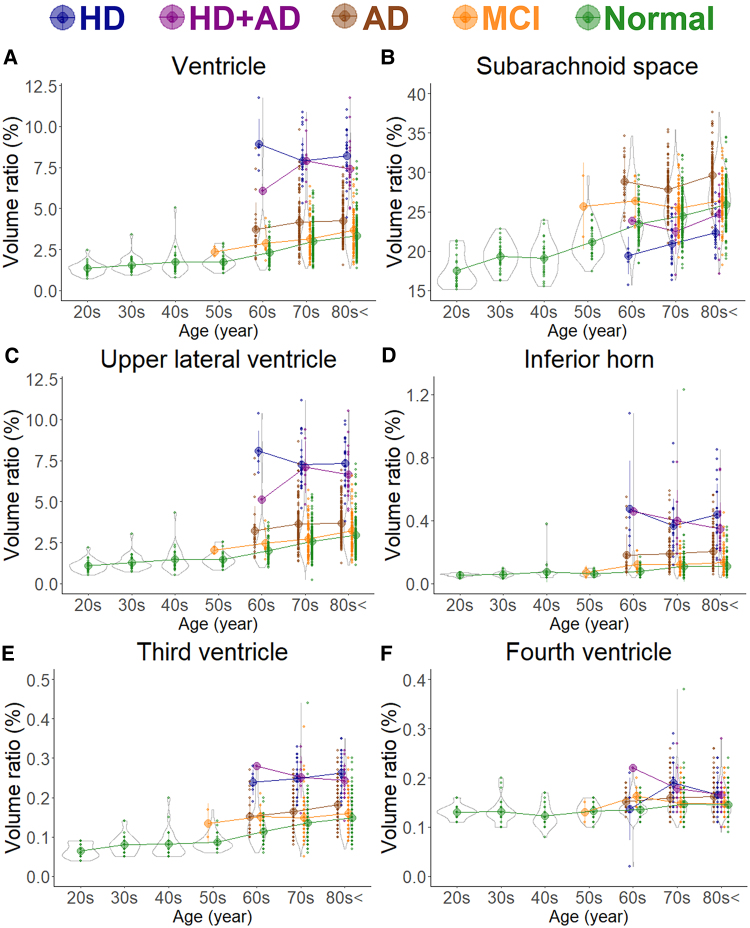
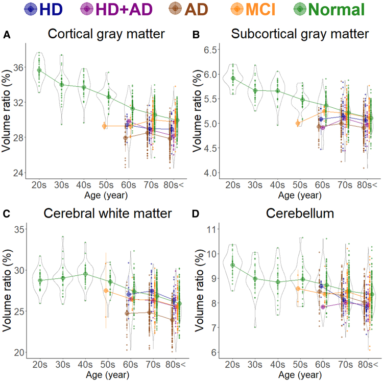
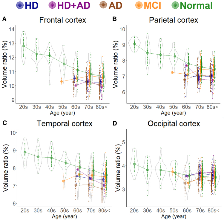
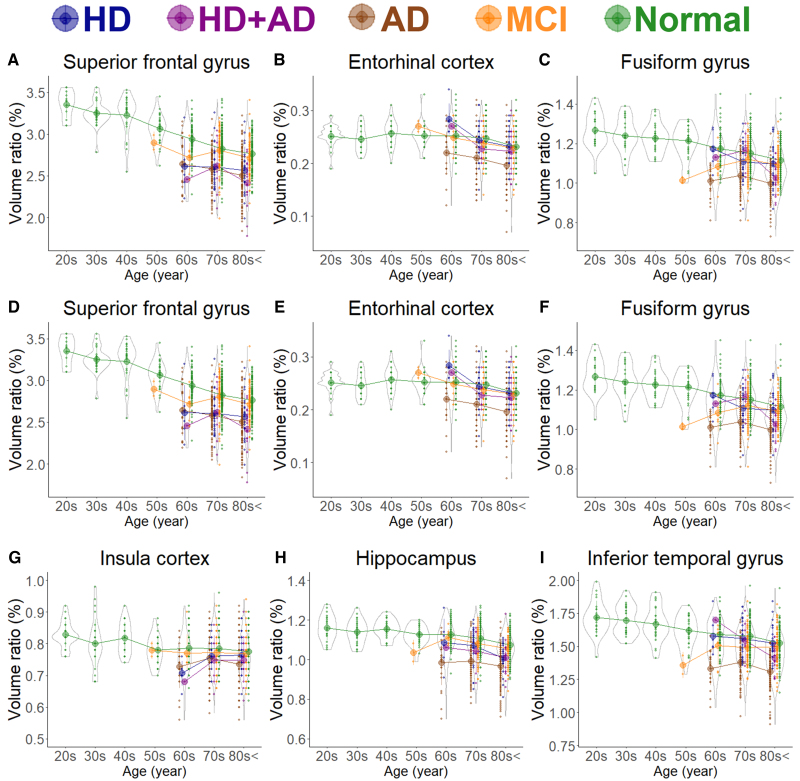
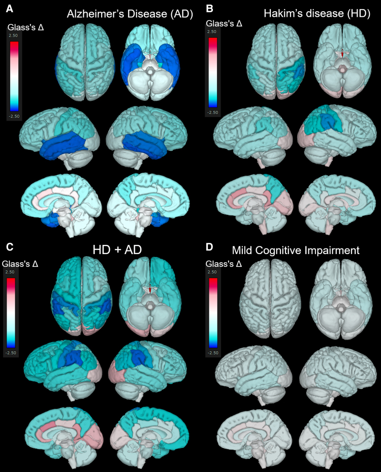
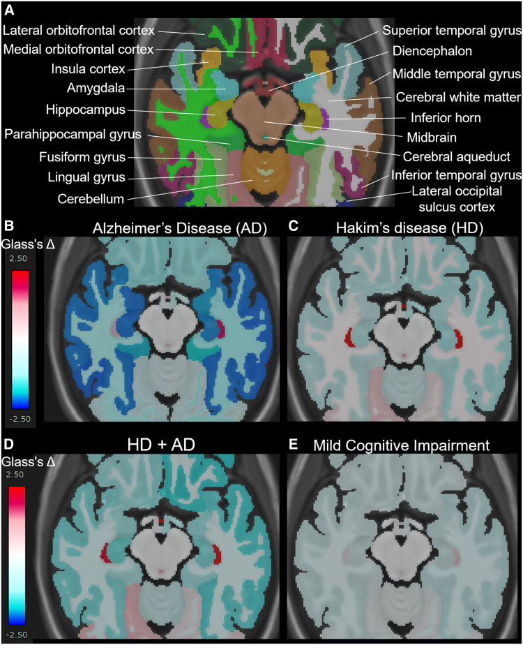
Idiopathic normal-pressure hydrocephalus (Hakim's disease) is characterized by ventricular enlargement and disproportionately enlarged subarachnoid space hydrocephalus, leading to localized brain deformation. Differentiating regional brain volume changes in Hakim's disease from those in Alzheimer's disease, Hakim's disease with Alzheimer's disease, and mild cognitive impairment provides insights into disease-specific mechanisms. This study aimed to identify disease-specific patterns of brain volume changes in Hakim's disease, Alzheimer's disease, Hakim's disease with Alzheimer's disease, and mild cognitive impairment and compare them with those in cognitively healthy individuals using an advanced artificial intelligence-based brain segmentation tool. The study included 970 participants, comprising 52 patients with Hakim's disease, 256 with Alzheimer's disease, 25 with Hakim's disease with Alzheimer's disease, 163 with mild cognitive impairment, and 474 healthy controls. The intracranial spaces were segmented into 100 brain and 7 CSF subregions from 3D T1-weighted MRIs using brain subregion analysis. The volume ratios of these regions were compared among the groups using Glass's Δ, referencing 400 healthy controls aged ≥50 years. Hakim's disease exhibited significant volume reduction in the supramarginal gyrus of the parietal lobe and the paracentral gyrus of the frontal lobe. Alzheimer's disease exhibited prominent volume loss in the hippocampus and temporal lobe, particularly in the entorhinal cortex, fusiform gyrus, and inferior temporal gyrus. Hakim's disease with Alzheimer's disease showed significant volume reductions in the supramarginal gyrus of the parietal lobe, similar to Hakim's disease, whereas temporal lobe volumes were relatively preserved compared with those in Alzheimer's disease. Patients with mild cognitive impairment aged ≥70 years had comparable regional brain volume ratios with healthy controls in the same age group. The Hakim's disease and Hakim's disease with Alzheimer's disease groups were characterized by volume reductions in the frontal and parietal lobes caused by disproportionately enlarged subarachnoid space hydrocephalus-related compression compared with temporal lobe atrophy observed in the Alzheimer's disease group. These disease-specific morphological changes highlight the need for longitudinal studies to clarify the causes of compression and atrophy.
Keywords: Alzheimer’s disease; Hakim’s disease; artificial intelligence; idiopathic normal-pressure hydrocephalus; mild cognitive impairment.
© The Author(s) 2025. Published by Oxford University Press on behalf of the Guarantors of Brain.
Conflict of interest statement
S.Y.amada has received a research grant from FUJIFILM Corporation and speaker honoraria from Fujifilm Medical Systems since 2019. T. Yuzawa and H. Ito are employed by FUJIFILM Corporation and have made substantial contributions to the development of the applications of the SYNAPSE 3D workstation. However, they were not involved in the results of this study, including data extraction or statistical analysis. The other authors of this manuscript have no relationships to declare with any of the companies whose products or services may be related to the subject matter of the study.
Figures

Similar articles
-
Modeling cerebrospinal fluid dynamics across the entire intracranial space through integration of four-dimensional flow and intravoxel incoherent motion magnetic resonance imaging.Fluids Barriers CNS. 2024 May 30;21(1):47. doi: 10.1186/s12987-024-00552-6. Fluids Barriers CNS. 2024. PMID: 38816737 Free PMC article.
-
Structural magnetic resonance imaging for the early diagnosis of dementia due to Alzheimer's disease in people with mild cognitive impairment.Cochrane Database Syst Rev. 2020 Mar 2;3(3):CD009628. doi: 10.1002/14651858.CD009628.pub2. Cochrane Database Syst Rev. 2020. PMID: 32119112 Free PMC article.
-
The brain as a sponge: a computed tomographic look at Hakim's hypothesis.Neurosurgery. 1984 Jun;14(6):670-5. doi: 10.1227/00006123-198406000-00004. Neurosurgery. 1984. PMID: 6462401
-
Gait Apraxia and Hakim's Disease: A Historical Review.Biomedicines. 2023 Apr 3;11(4):1086. doi: 10.3390/biomedicines11041086. Biomedicines. 2023. PMID: 37189704 Free PMC article. Review.
-
Changes in the Parietal Lobe Subregion Volume at Various Stages of Alzheimer's Disease and the Role in Cognitively Normal and Mild Cognitive Impairment Conversion.J Integr Neurosci. 2025 Jan 21;24(1):25991. doi: 10.31083/JIN25991. J Integr Neurosci. 2025. PMID: 39862009
References
-
- Adams RD, Fisher CM, Hakim S, Ojemann RG, Sweet WH. Symptomatic occult hydrocephalus with “normal” cerebrospinal-fluid pressure. A treatable syndrome. N Engl J Med. 1965;273:117–126. - PubMed
-
- Marmarou A, Bergsneider M, Relkin N, Klinge P, Black PM.. Development of guidelines for idiopathic normal-pressure hydrocephalus: Introduction. Neurosurgery. 2005;57(3 Suppl):S1–S3; discussion ii-v. - PubMed
-
- Relkin N, Marmarou A, Klinge P, Bergsneider M, Black PM.. Diagnosing idiopathic normal-pressure hydrocephalus. Neurosurgery. 2005;57(3 Suppl):S4–S16; discussion ii-v. - PubMed
-
- Ishikawa M; Guideline Committe for Idiopathic Normal Pressure Hydrocephalus JSoNPH . Clinical guidelines for idiopathic normal pressure hydrocephalus. Neurol Med Chir (Tokyo). 2004;44(4):222–223. - PubMed
-
- Marmarou A, Black P, Bergsneider M, Klinge P, Relkin N; International NPH Consultant Group . Guidelines for management of idiopathic normal pressure hydrocephalus: Progress to date. Acta Neurochir Suppl. 2005;95:237–240. - PubMed
LinkOut - more resources
Full Text Sources