[Preprint]. 2025 May 5:2024.09.27.615498.
doi: 10.1101/2024.09.27.615498.

Genome-directed study reveals the diversity of Salmonella T6SS effectors and identifies a novel family of lipid-targeting antibacterial toxins

Affiliations

Genome-directed study reveals the diversity of Salmonella T6SS effectors and identifies a novel family of lipid-targeting antibacterial toxins

Gianlucca G Nicastro et al. bioRxiv. .

Abstract

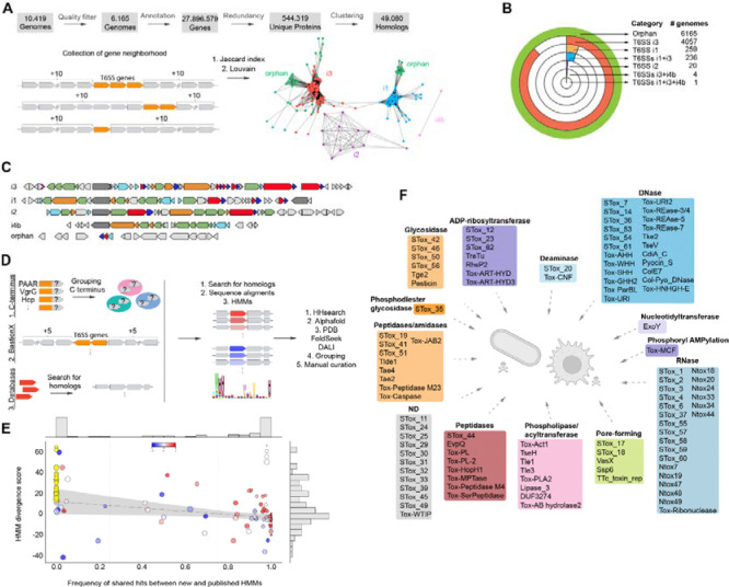
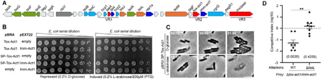
Bacterial warfare is a common and ancient phenomenon in nature, where bacterial species use strategies to inhibit the growth or kill competitors. This involves the production and deployment of antibacterial toxins that disrupt essential cellular processes in target cells. The continuous arms race in which bacteria acquire new toxin and immunity proteins to promote increased adaptation to their environment is responsible for the diversification of this toxin repertoire. Here, we deployed in-silico strategies to analyze 10,000 genomes and identify effectors secreted via the type VI secretion system of Salmonella. We identified 128 candidates, which are widespread in a vast array of Salmonella serovars and other bacterial species. Tox-Act1 is among the most frequent candidates and was selected for in-depth characterization. Tox-Act1 contains a permuted NlpC/P60 papain-like catalytic core characteristic of lipid-targeting members rather than the typical peptidases or amidases. Evolutionary analysis revealed the relationship of Tox-Act1 with acyltransferases. Biochemical assays with purified toxin and lipidomics of intoxicated cells showed that Tox-Act1 exhibits phospholipase activity, cleaving off acyl groups from phosphatidylglycerol and phosphatidylethanolamine. In addition, we demonstrate that Tox-Act1 is secreted in a T6SS-dependent manner and provide a competitive advantage during colonization of the gut of infected mice. This work broadens our understanding of toxin domains and provides the first direct characterization of a lipid-targeting NlpC/P60 domain in biological conflicts.

Keywords: NlpC/P60; Salmonella; T6SS; acyltransferase; effector; phospholipase; toxin.


Figures

Fig 1.
Fig 1.. Computational pipeline for the identification of T6SS subtypes and effector repertoire within 10,000 Salmonella genomes.
(a) Pipeline used for classification of genomic sites and T6SS subtypes. (b) Number of genomes containing the different T6SS subtypes within the 10KSG dataset. (c) Examples of the genomic organization of T6SS structural clusters from distinct phylogenetic subtypes. Colors denote structural proteins forming the membrane complex (orange), sheath and inner tube (light blue), baseplate and spike components (green). Effectors are shown in red and immunities in dark blue. (d) In silico strategies used for the identification and classification of T6SS effectors. (e) Comparison of profile Hidden Markov Models (pHMMs) of STox to published models of related families. Each circle corresponds to a population of proteins detected by an STox model. The relative frequency of proteins detected by an STox model compared to a reference model is shown on the horizontal axis. The HMM divergence score is shown in the vertical axis. The blue-white-red color scale of each dot represents the values for the Spearman correlation index between STox and reference alignment scores for the same proteins. Yellow circles represent STox models that detect proteins that are not recognized by previously existing models. The radius of each circle is proportional to the total number of proteins detected only by STox models. The regression line does not include the data points for the models represented by yellow circles. (f) Schematic representation of the functional classes of T6SS effectors identified in the 10KSG.
Fig 2.
Fig 2.. Unique subsets of effectors are associated with specific Salmonella serovars and T6SS subtypes.
(a) The most frequent effectors identified in the 10KSG dataset. Each bar represents the number of genomes encoding a specific effector. Colors represent different effector activities, with light colors representing orphan effectors while dark colors represent effectors encoded within the structural cluster. (b) Schematic representation of the most common sets of effectors in genomes encoding different T6SS subtypes. The number of genomes is indicated on the right. Colors represent activity as shown in (a). (c) The five most frequent effectors encoded in different Salmonella serovars. Colors indicate the effector activity as in (a). (d) Pie chart illustrating the relative proportions of effectors classified by activity encoded within the T6SSs subtypes i3, i1, and i2. Colors as shown in (a). (e) Venn diagram illustrating the proportion of overlap between effectors encoded within each T6SS structural cluster (blue: i1; purple: subtype i2; red: subtype i3; and green: orphan). (f) Schematic representation of the genetic organization of T6SSs showing the position of variable regions in which the effector and immunity proteins are encoded. Colors denote structural proteins forming the membrane complex (orange), sheath and inner tube (light blue), baseplate and spike components (green). Effectors are shown in red, and immunities in dark blue.
Fig 3.
Fig 3.. Tox-Act1 is a T6SS effector used for bacterial competition in the mouse gut.
(a) Scheme of the genomic region encoding Tox-Act1 and Imm-Act1 effector/immunity pair (FD01843896). (b) E. coli toxicity assay. Serial dilutions of E. coli carrying pBRA and pEXT22 constructs. Images are representative of thee independent experiments. (c) Time-lapse microscopy of E. coli carrying pBRA SP-Tox-Act1 grown on repressed or induced conditions. Scale bar: 5 μm. Timestamps in hh:mm. (d) C57BL/6 mice were infected by oral gavage with equal numbers of each strain. Bacteria were recovered from the cecum 4 days after infection, and CI values calculated. The log10 Cis were used for statistical analysis. Single sample t-test was used to compare the CI to the hypothetical value of 0, and p value is indicated in brackets. Unpaired t-test (**p <0.01) was used to compare the two groups.
Fig 4.
Fig 4.. Tox-Act1 is evolutionarily related to lipid-targeting enzymes with a permuted NlpC/P60 domain.
(a) Maximum-likelihood phylogenetic tree of permuted NlpC/P60 members. Dots represent the number of PSI-BLAST iterations required to collect homologs and the red star marks the query. (b) Genomic organization of representatives from clades TseH and Tox-Act1 showing the genes are encoded in the context of conflict systems, and DUF4105 showing context of lipid metabolism. (c) Sequence logo from the enzymatic core of permuted NlpC/P60 from all clades shown in (a). The arrows indicate conserved His and Cys residues that were mutated in (d). (d) E. coli toxicity assay. Serial dilution of E. coli containing pBRA and pEXT22 constructs. Images are representative of thee independent experiments.
Fig 5.
Fig 5.. Tox-Act1 has phospholipase A activity and changes the composition of target cell membranes.
(a) In vitro enzymatic assay with recombinant Tox-Act1 (red) or Tox-Act1C151A (blue) incubated with different phospholipids (both 16:0–18:1 PG and PE). The amount of lysophospholipids produced was analyzed and quantified by HPLC-MS/MS. (b) Quantification of the peak area of lysophospholipids normalized by the intact substrate. Data corresponds to the mean ± SD. ***p < 0.001 and *p < 0.05, ns not significant (unpaired t-test). (c) UHPLC-MS total ion chomatogram showing the profile of total lipids extracted from E. coli expressing Tox-Act1 (red) or Tox-Act1C151A (blue). (d) Heatmap plot of top 20 altered lipids of intoxicated E. coli. Results display four biological replicates of each condition (WT or C151A) with the quantification of lipids species (Tukey test; p < 0.05 FDR adjusted). Data were expressed in nM mg of protein−1 and normalized by log transformation (base 10) prior to analysis. Red (up) and blue (down) bars represent changes in lipid species concentration relative to the normalized mean. Letters differentiate between the isomers.

Similar articles

References

    1. Granato ET, Meiller-Legrand TA, Foster KR. The Evolution and Ecology of Bacterial Warfare. Curr Biol. 2019;29(11):R521–R37. doi: 10.1016/j.cub.2019.04.024. - DOI - PubMed
    1. Hood RD, Peterson SB, Mougous JD. From Striking Out to Striking Gold: Discovering that Type VI Secretion Targets Bacteria. Cell host & microbe. 2017;21(3):286–9. doi: 10.1016/j.chom.2017.02.001. - DOI - PMC - PubMed
    1. Russell AB, Wexler AG, Harding BN, Whitney JC, Bohn AJ, Goo YA, et al. A type VI secretion-related pathway in Bacteroidetes mediates interbacterial antagonism. Cell host & microbe. 2014;16(2):227–36. Epub 20140725. doi: 10.1016/j.chom.2014.07.007. - DOI - PMC - PubMed
    1. Li J, Yao Y, Xu HH, Hao L, Deng Z, Rajakumar K, et al. SecReT6: a web-based resource for type VI secretion systems found in bacteria. Environ Microbiol. 2015;17(7):2196–202. doi: 10.1111/1462-2920.12794. - DOI - PubMed
    1. Bock D, Medeiros JM, Tsao HF, Penz T, Weiss GL, Aistleitner K, et al. In situ architecture, function, and evolution of a contractile injection system. Science. 2017;357(6352):713–7. doi: 10.1126/science.aan7904. - DOI - PMC - PubMed

Publication types

LinkOut - more resources