. 2025 Mar 3;9(2):102720.
doi: 10.1016/j.rpth.2025.102720. eCollection 2025 Feb.

Factor XI localization in human deep venous thrombus and function of activated factor XI on venous thrombus formation and hemostasis

Affiliations

Factor XI localization in human deep venous thrombus and function of activated factor XI on venous thrombus formation and hemostasis

Nobuyuki Oguri et al. Res Pract Thromb Haemost. .

Abstract

Background: Novel anticoagulants targeting coagulation factor (F)XI/activated FXI (FXIa) are currently under development. However, whether FXI is present in human deep vein thrombosis (DVT) and whether FXIa and activated FX (FXa) play different roles in venous thrombus formation and hemostasis remain unclear.

Objectives: To determine the presence of FXI in DVT and the effects of direct oral FXIa and FXa inhibitors on venous thrombus formation and hemostasis in rabbits and on in vitro thrombus formation.

Methods: We immunohistochemically assessed FXI localization in human-aspirated DVT (n = 15). Additionally, we compared thrombus formation induced by endothelial denudation and stenosis or stasis in the jugular vein and skin bleeding time and volume between rabbits treated with direct FXIa inhibitors (ONO-1600586) and FXa inhibitors (rivaroxaban). Ex vivo rabbit and human blood were perfused in a flow chamber under low-shear rates (70/s).

Results: FXI was localized in all DVT, predominantly in fibrin-rich areas. The FXI immunopositive area in the nonorganizing area was greater than that in the organizing area. Although FXIa and FXa inhibitors comparably inhibited venous thrombus formation, FXIa inhibitors did not affect bleeding time or volume in rabbits. FXIa or FXa inhibitors mildly or strongly inhibited fibrin formation at low-shear rates, respectively. Furthermore, the FXIa inhibitor suppressed human FXIa activity, thrombin generation, and fibrin formation during perfusion.

Conclusion: The pathologic findings of human DVT suggest FXI's role in human DVT. FXIa inhibitors may inhibit less fibrin formation than FXa inhibitors and may explain the minor role of FXIa in hemostasis.

Keywords: factor X; factor XI; hemostasis; pathology; venous thromboembolism.


Figures

None
Graphical abstract
Figure 1
Figure 1
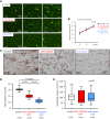
Presence of factor (F)XI in human deep vein thrombosis (DVT). (A) Representative immunofluorescent images of fresh components of DVT. The upper row shows CF488-labeled FXI (green), CF568-labeled fibrin (red), and merged images. The second row shows CF488-labeled FXI, CF568-labeled glycophorin A, and merged images. The third row shows CF488-labeled FXI, CF568-labeled CD42b, and merged images. The lower row shows CF488-labeled FXI, CF568-labeled CD66b, and merged images. FXI was closely localized with fibrin. Glycophorin A, a marker of erythrocytes, was predominantly surrounded by FXI. FXI was partly localized with CD42b, a marker of platelets, but hardly localized with CD66b, a marker of neutrophils. (B) Representative immunofluorescent images of fresh components of DVT show CF488-labeled FXI, CF568-labeled FXII, 4,6-diamidino-2-phenylindole (DAPI, a DNA marker), and merged images. FXI was partly localized with DNA and FXII. (C) Representative images of in situ nanogold labeling and electron microscopy of fresh components of DVT. The left upper image shows an immunohistochemical image of FXI. Nanogolds accumulate at the 3, 3′-diaminobenzidine tetrahydrochloride deposition site against anti-FXI antibody (left lower). Nanogold deposition was localized on a fibrin-like mesh network but not on the neutrophils (arrow). (D) Representative immunohistochemical images in fresh and organizing areas of DVT. The organizing areas were confirmed by the presence of CD34-immunopositive cells, corresponding to endothelialization/organization, while the fresh area was confirmed by the absence of CD34-immunopositive cells. FXI localized fibrin- and erythrocyte-rich fresh areas. In the fresh area, aggregated clusters of platelets and sparsely distributed neutrophils are present. The inset in CD66b represents a high-magnification image of the dashed square. In the organizing area, immunoreaction for FXI, fibrin, glycoprotein (GP) IIb/IIIa, glycophorin A, or CD66b is modest or focal. (E) Immunopositive area for FXI, fibrin, GPIIb/IIIa, and glycophorin A, and CD66b-immunopositive cell density in nonorganizing (CD34-immunonegative) and organizing (CD34-immunopositive) areas (Mann–Whitney U-test). HE, hematoxylin and eosin.
Figure 1
Figure 1
Presence of factor (F)XI in human deep vein thrombosis (DVT). (A) Representative immunofluorescent images of fresh components of DVT. The upper row shows CF488-labeled FXI (green), CF568-labeled fibrin (red), and merged images. The second row shows CF488-labeled FXI, CF568-labeled glycophorin A, and merged images. The third row shows CF488-labeled FXI, CF568-labeled CD42b, and merged images. The lower row shows CF488-labeled FXI, CF568-labeled CD66b, and merged images. FXI was closely localized with fibrin. Glycophorin A, a marker of erythrocytes, was predominantly surrounded by FXI. FXI was partly localized with CD42b, a marker of platelets, but hardly localized with CD66b, a marker of neutrophils. (B) Representative immunofluorescent images of fresh components of DVT show CF488-labeled FXI, CF568-labeled FXII, 4,6-diamidino-2-phenylindole (DAPI, a DNA marker), and merged images. FXI was partly localized with DNA and FXII. (C) Representative images of in situ nanogold labeling and electron microscopy of fresh components of DVT. The left upper image shows an immunohistochemical image of FXI. Nanogolds accumulate at the 3, 3′-diaminobenzidine tetrahydrochloride deposition site against anti-FXI antibody (left lower). Nanogold deposition was localized on a fibrin-like mesh network but not on the neutrophils (arrow). (D) Representative immunohistochemical images in fresh and organizing areas of DVT. The organizing areas were confirmed by the presence of CD34-immunopositive cells, corresponding to endothelialization/organization, while the fresh area was confirmed by the absence of CD34-immunopositive cells. FXI localized fibrin- and erythrocyte-rich fresh areas. In the fresh area, aggregated clusters of platelets and sparsely distributed neutrophils are present. The inset in CD66b represents a high-magnification image of the dashed square. In the organizing area, immunoreaction for FXI, fibrin, glycoprotein (GP) IIb/IIIa, glycophorin A, or CD66b is modest or focal. (E) Immunopositive area for FXI, fibrin, GPIIb/IIIa, and glycophorin A, and CD66b-immunopositive cell density in nonorganizing (CD34-immunonegative) and organizing (CD34-immunopositive) areas (Mann–Whitney U-test). HE, hematoxylin and eosin.
Figure 2
Figure 2
Activated partial thromboplastin time (aPTT) and plasma prothrombin time (PT) before and after oral administration of ONO-1600586 (activated factor [F]XI inhibitor) and rivaroxaban (activated FX inhibitor). (A) aPTT before and 75 minutes (before thrombus formation and bleeding test) and 4.5 hours (3 hours after thrombus formation) after oral administration of the solvent (control), ONO-1600586, and rivaroxaban (Kruskal–Wallis test with Dunn’s multiple comparisons test). aPTT before administration showed significant differences among the groups. However, there was no significant difference between the control and others (FXIa and FXa inhibitors) or between each group and every other group in Dunn’s multiple comparison test; n means the number of animals. (B) PT before and 75 minutes (before thrombus formation and bleeding test) and 4.5 hours (3 hours after thrombus formation) after oral administration of the control, ONO-1600586, and rivaroxaban (Kruskal–Wallis test with Dunn’s multiple comparisons test; n means the number of animals).
Figure 3
Figure 3
Different contributions of activated factor (F)XI and activated FX to venous thrombus formation with endothelial denudation and luminal stenosis and skin bleeding in rabbits. (A) Representative macroscopic images of the postfixed venous thrombi. Thrombi are dark reddish in color in each group. Thrombi of rivaroxaban and ONO-1600586 administration groups are smaller than those of the control (solvent) group. (B) Weight of unfixed venous thrombus in control, ONO-1600586, or rivaroxaban administration groups. Oral administration of ONO-1600586 (50 mg/kg) and rivaroxaban (15 mg/kg) similarly reduced venous thrombus weight compared with that of the control. Each group was compared with the control group (Kruskal–Wallis test with Dunn’s multiple comparison test; n means the number of animals). (C) Representative histologic and immunohistochemical images of venous thrombus in control, ONO-1600586, or rivaroxaban groups. All thrombi are rich in erythrocytes and contain fibrin and platelets (glycoprotein [GP] IIb/IIIa). (D) The areas of erythrocytes or immunopositive areas for fibrin or GPIIb/IIIa in venous thrombus (Kruskal–Wallis test; n means the number of histologic sections). (E) Bleeding time and bleeding volume after oral administration of the control, ONO-1600586, and rivaroxaban. Rivaroxaban dose-dependently prolonged bleeding time (left) and increased bleeding volume (right). Oral administration of ONO-1600586 did not affect bleeding time or volume. Each group was compared with the control group (Kruskal–Wallis test with Dunn’s multiple comparisons test; n means the number of histologic sections). HE, hematoxylin and eosin.
Figure 4
Figure 4
Weight of unfixed stasis-induced venous thrombus in rabbits. Oral administration of ONO-1600586 (50 mg/kg) and rivaroxaban (15 mg/kg) similarly reduced venous thrombus weight compared with that of the control (Kruskal–Wallis test with Dunn’s multiple comparison test; n means the number of animals).
Figure 5
Figure 5
Contributions of activated factor (F)XI and activated FX to ex vivo thrombus formation in a flow chamber system. Rabbit blood was collected 75 minutes after oral administration of a solvent (control), ONO-1600586 (50 mg/kg), and rivaroxaban (15 mg/kg). The ex vivo blood was perfused in a flow chamber under a low-shear rate of 70/s. (A) Representative fluorescent images under a low-shear rate (70/s). Platelets and leukocytes were labeled with mepacrine, and the fluorescent images were captured 3, 6, and 9 minutes after perfusion. The surface covering area time-dependently increased in all 3 groups. Large fluorescent dots indicate leukocytes (arrows). (B) The surface covering areas under low-shear rate in 3 groups (2-way repeated measure anova with Tukey’s multiple comparison test). (C) Representative immunostaining images of fibrin on the glass coverslips after perfusion. The mesh-like pattern of fibrin formation on islands of aggregated platelets and between the islands in the control. Reduced fibrin formation in ONO-1600586 administration. Absence or little fibrin formation in rivaroxaban administration. (D) Fibrin-immunopositive area on the glass coverslips after ex vivo blood perfusion under low-shear rate (Kruskal–Wallis test with Dunn’s multiple comparison test). (E) The number of leukocyte adhesions after ex vivo blood perfusion under low-shear rate (Kruskal–Wallis test).
Figure 6
Figure 6
Effects of ONO-1600586 on human activated factor (F)XI (FXIa) and in vitro thrombus formation. The human blood was perfused in a flow chamber under a low-shear rate of 70/s. (A) Representative fluorescent images under a low-shear rate (70/s). Platelets and leukocytes were labeled with mepacrine, and the fluorescent images were captured 3, 6, and 9 minutes after perfusion. The surface covering area time-dependently increased in all 3 groups. Large fluorescent dots indicate leukocytes (arrows). (B) The surface covering areas under a low-shear rate in the control (solvent), ONO-1600586, and rivaroxaban addition (2-way repeated measure anova with Tukey’s multiple comparisons test). (C) Representative immunostaining images of fibrin on the glass coverslips after perfusion. ONO-1600586 or rivaroxaban addition decreased fibrin formation. (D) Fibrin-immunopositive area after human blood perfusion under a low-shear rate (Kruskal–Wallis test with Dunn’s multiple comparison test). (E) The number of leukocyte adhesions after human blood perfusion under a low-shear rate (Kruskal–Wallis test). (F) Activated FX activity, FXIa activity, and levels of prothrombin fragment 1 + 2 before and after perfusion of human blood (2-way repeated measure anova with Tukey’s multiple comparison test). ∗P < .0001 vs control; †P < .0001 vs ONO-1600586; ‡P < .01 vs ONO-1600586. (G) Representative immunofluorescent images of the glass coverslips after perfusion. The upper row shows CF488-labeled FXI (green), CF568-labeled fibrin (red), and merged images. The second row shows CF488-labeled FXI, CD42b, and merged images. The lower row shows CF488-labeled FXI, CF568-labeled CD66b, and merged images. FXI was closely localized with fibrin and partly around CD42b, a marker of platelets, but hardly localized with CD66b, a marker of neutrophils.
Figure 6
Figure 6
Effects of ONO-1600586 on human activated factor (F)XI (FXIa) and in vitro thrombus formation. The human blood was perfused in a flow chamber under a low-shear rate of 70/s. (A) Representative fluorescent images under a low-shear rate (70/s). Platelets and leukocytes were labeled with mepacrine, and the fluorescent images were captured 3, 6, and 9 minutes after perfusion. The surface covering area time-dependently increased in all 3 groups. Large fluorescent dots indicate leukocytes (arrows). (B) The surface covering areas under a low-shear rate in the control (solvent), ONO-1600586, and rivaroxaban addition (2-way repeated measure anova with Tukey’s multiple comparisons test). (C) Representative immunostaining images of fibrin on the glass coverslips after perfusion. ONO-1600586 or rivaroxaban addition decreased fibrin formation. (D) Fibrin-immunopositive area after human blood perfusion under a low-shear rate (Kruskal–Wallis test with Dunn’s multiple comparison test). (E) The number of leukocyte adhesions after human blood perfusion under a low-shear rate (Kruskal–Wallis test). (F) Activated FX activity, FXIa activity, and levels of prothrombin fragment 1 + 2 before and after perfusion of human blood (2-way repeated measure anova with Tukey’s multiple comparison test). ∗P < .0001 vs control; †P < .0001 vs ONO-1600586; ‡P < .01 vs ONO-1600586. (G) Representative immunofluorescent images of the glass coverslips after perfusion. The upper row shows CF488-labeled FXI (green), CF568-labeled fibrin (red), and merged images. The second row shows CF488-labeled FXI, CD42b, and merged images. The lower row shows CF488-labeled FXI, CF568-labeled CD66b, and merged images. FXI was closely localized with fibrin and partly around CD42b, a marker of platelets, but hardly localized with CD66b, a marker of neutrophils.

References

    1. Wendelboe A.M., Raskob G.E. Global burden of thrombosis: epidemiologic aspects. Circ Res. 2016;118:1340–1347. - PubMed
    1. Jiang H., Jiang Y., Ma H., Zeng H., Lv J. Effects of rivaroxaban and warfarin on the risk of gastrointestinal bleeding and intracranial hemorrhage in patients with atrial fibrillation: systematic review and meta-analysis. Clin Cardiol. 2021;44:1208–1215. - PMC - PubMed
    1. Carnicelli A.P., Hong H., Connolly S.J., Eikelboom J., Giugliano R.P., Morrow D.A., et al. Direct oral anticoagulants versus warfarin in patients with atrial fibrillation: patient-level network meta-analyses of randomized clinical trials with interaction testing by age and sex. Circulation. 2022;145:242–255. - PMC - PubMed
    1. Gailani D., Broze G.J., Jr. Factor XI activation in a revised model of blood coagulation. Science. 1991;253:909–912. - PubMed
    1. Meijers J.C., Tekelenburg W.L., Bouma B.N., Bertina R.M., Rosendaal F.R. High levels of coagulation factor XI as a risk factor for venous thrombosis. N Engl J Med. 2000;342:696–701. - PubMed

LinkOut - more resources